

Effects of nimesulide on the small intestine mucositis induced by methotrexate in rats
- PMID: 27333839
- PMCID: PMC5111835
- DOI: 10.1538/expanim.15-0122
Effects of nimesulide on the small intestine mucositis induced by methotrexate in rats
Abstract
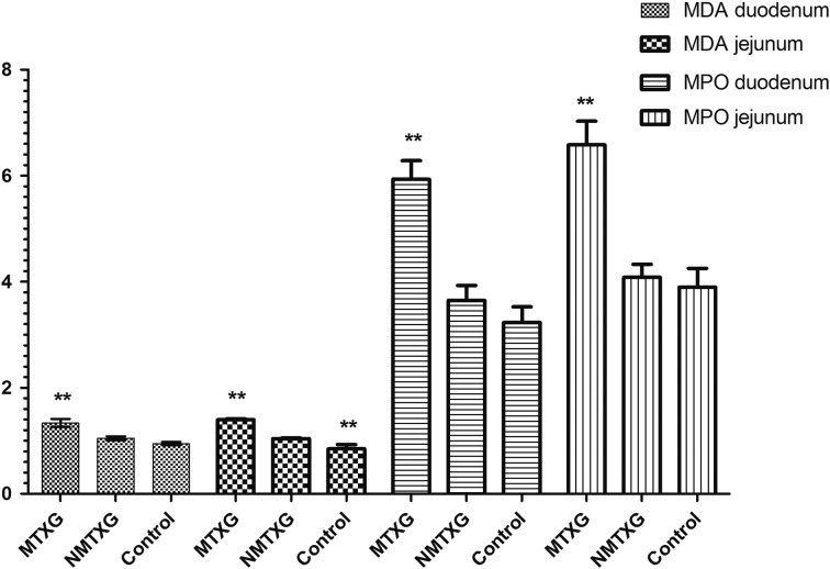
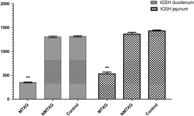
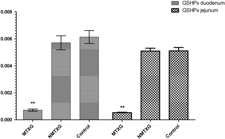
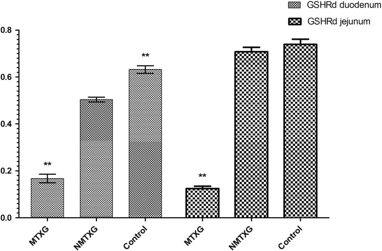
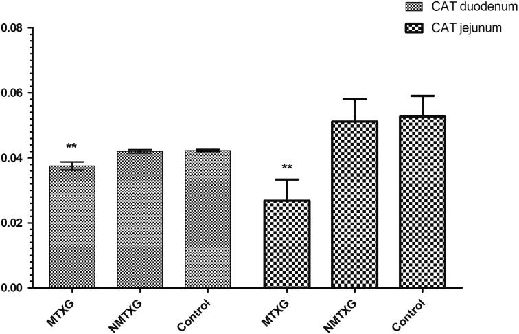
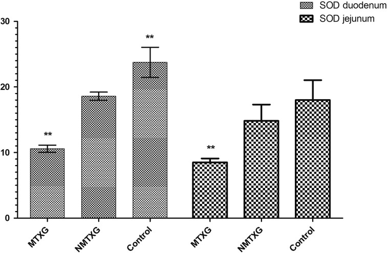
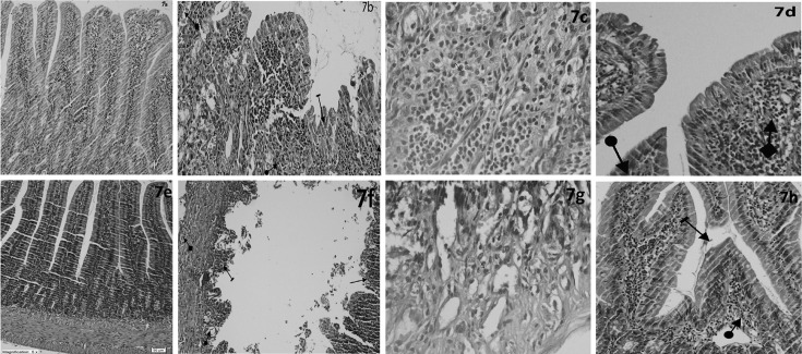
Intestinal mucositis is one of the major problems in the patients receiving cancer treatment. Nimesulide is a drug with antioxidant, antiinflammatory and antiulcer features. We aimed to investigate the effect of nimesulide on the small intestine mucositis induced by methotrexate (MTX) in rats. Experimental animals were divided into the control group, MTX group (MTXG) and nimesulide+MTX administered group (NMTXG) with eight rats per group. The control and MTXG groups were given distilled water by gavage and the NMTXG was given nimesulide 100 mg/kg orally. After one hour, the NMTXG and MTXG rat groups were administered oral MTX 5 mg/kg. This procedure was repeated once a day for 15 days and the rats were sacrificed. The duodenum and jejunum of each rat was removed for the assessment of biochemical markers and histopathological evaluation. Malondialdehyde (MDA) and myeloperoxidase (MPO) levels were significantly higher in the duodenal and jejunal tissues of the animals which received MTX, compared to the control and NMTXG (P<0.001). Also, the levels of total glutathione (tGSH), glutathione reductase (GSHRd), glutathione peroxidase (GSHPx), catalase (CAT) and superoxide dismutase (SOD) were significantly lower in the MTXG (P<0.001) compared to other groups. MTX led to villus and crypt epithelial damage and inflammation containing marked PMNL and eosinophils in the intestinal tissues histopathologically. Whereas, there was only mild irregularities in the villus structures of the NMTXG. Nimesulide protected the small intestines against damage by MTX. Intestinal mucositis caused by MTX may be preventable by co-administered nimesulide.
Figures
Similar articles
-
Effects of anakinra on the small intestine mucositis induced by methotrexate in rats.Exp Anim. 2020 Apr 24;69(2):144-152. doi: 10.1538/expanim.19-0057. Epub 2019 Dec 2. Exp Anim. 2020. PMID: 31787709 Free PMC article.
-
Efficacy of royal jelly on methotrexate-induced systemic oxidative stress and damage to small intestine in rats.Afr J Tradit Complement Altern Med. 2012 Apr 2;9(3):412-7. doi: 10.4314/ajtcam.v9i3.17. eCollection 2012. Afr J Tradit Complement Altern Med. 2012. PMID: 23983375 Free PMC article.
-
In a methotrexate-induced model of intestinal mucositis, olmesartan reduced inflammation and induced enteropathy characterized by severe diarrhea, weight loss, and reduced sucrose activity.Biol Pharm Bull. 2015;38(5):746-52. doi: 10.1248/bpb.b14-00847. Biol Pharm Bull. 2015. PMID: 25947920
-
Steamed root of Rehmannia glutinosa Libosch (Plantaginaceae) alleviates methotrexate-induced intestinal mucositis in rats.J Ethnopharmacol. 2016 May 13;183:143-150. doi: 10.1016/j.jep.2016.02.035. Epub 2016 Mar 2. J Ethnopharmacol. 2016. PMID: 26934449
-
Mucositis and non-invasive markers of small intestinal function.Cancer Biol Ther. 2009 May;8(9):753-8. doi: 10.4161/cbt.8.9.8232. Epub 2009 May 19. Cancer Biol Ther. 2009. PMID: 19276675 Review.
Cited by
-
Methotrexate Ameliorates Systemic Inflammation and Septic Associated-Lung Damage in a Cecal Ligation and Puncture Septic Rat Model.Int J Mol Sci. 2021 Sep 4;22(17):9612. doi: 10.3390/ijms22179612. Int J Mol Sci. 2021. PMID: 34502521 Free PMC article.
-
Effects of anakinra on the small intestine mucositis induced by methotrexate in rats.Exp Anim. 2020 Apr 24;69(2):144-152. doi: 10.1538/expanim.19-0057. Epub 2019 Dec 2. Exp Anim. 2020. PMID: 31787709 Free PMC article.
-
Experimental Chemotherapy-Induced Mucositis: A Scoping Review Guiding the Design of Suitable Preclinical Models.Int J Mol Sci. 2022 Dec 6;23(23):15434. doi: 10.3390/ijms232315434. Int J Mol Sci. 2022. PMID: 36499758 Free PMC article.
References
-
- Beutheu S., Ouelaa W., Guérin C., Belmonte L., Aziz M., Tennoune N., Bôle-Feysot C., Galas L., Déchelotte P., Coëffier M.2014. Glutamine supplementation, but not combined glutamine and arginine supplementation, improves gut barrier function during chemotherapy-induced intestinal mucositis in rats. Clin. Nutr. 33: 694–701. doi: 10.1016/j.clnu.2013.09.003 - DOI - PubMed
-
- Beutler E.1971. Red cell metabolism: a manual of biochemical methods. Academic Press, London.
Publication types
MeSH terms
Substances
LinkOut - more resources
Full Text Sources
Other Literature Sources
Research Materials
Miscellaneous