. 2016 Jun 22;13(1):161.
doi: 10.1186/s12974-016-0611-x.

Complement is activated in progressive multiple sclerosis cortical grey matter lesions

Affiliations

Complement is activated in progressive multiple sclerosis cortical grey matter lesions

Lewis M Watkins et al. J Neuroinflammation. .

Abstract

Background: The symptoms of multiple sclerosis (MS) are caused by damage to myelin and nerve cells in the brain and spinal cord. Inflammation is tightly linked with neurodegeneration, and it is the accumulation of neurodegeneration that underlies increasing neurological disability in progressive MS. Determining pathological mechanisms at play in MS grey matter is therefore a key to our understanding of disease progression.

Methods: We analysed complement expression and activation by immunocytochemistry and in situ hybridisation in frozen or formalin-fixed paraffin-embedded post-mortem tissue blocks from 22 progressive MS cases and made comparisons to inflammatory central nervous system disease and non-neurological disease controls.

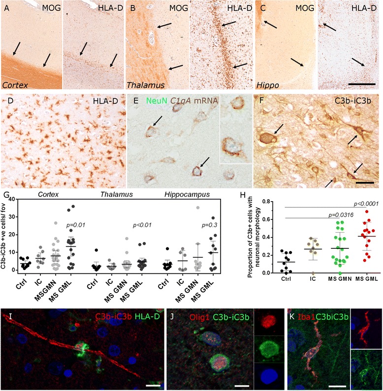
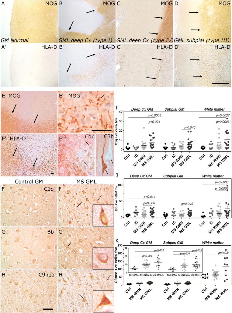
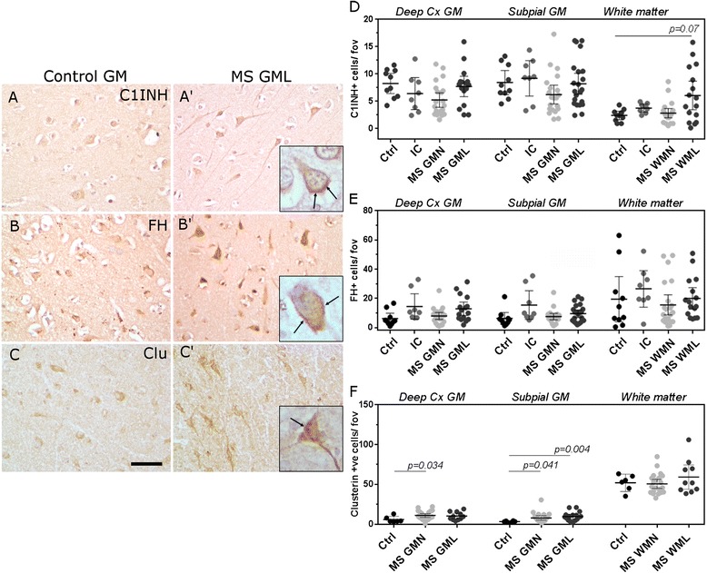
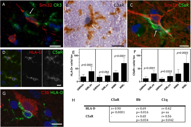
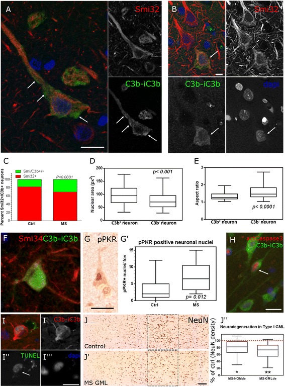
Results: Expression of the transcript for C1qA was noted in neurons and the activation fragment and opsonin C3b-labelled neurons and glia in the MS cortical and deep grey matter. The density of immunostained cells positive for the classical complement pathway protein C1q and the alternative complement pathway activation fragment Bb was significantly increased in cortical grey matter lesions in comparison to control grey matter. The number of cells immunostained for the membrane attack complex was elevated in cortical lesions, indicating complement activation to completion. The numbers of classical (C1-inhibitor) and alternative (factor H) pathway regulator-positive cells were unchanged between MS and controls, whilst complement anaphylatoxin receptor-bearing microglia in the MS cortex were found closely apposed to cortical neurons. Complement immunopositive neurons displayed an altered nuclear morphology, indicative of cell stress/damage, supporting our finding of significant neurodegeneration in cortical grey matter lesions.

Conclusions: Complement is activated in the MS cortical grey matter lesions in areas of elevated numbers of complement receptor-positive microglia and suggests that complement over-activation may contribute to the worsening pathology that underlies the irreversible progression of MS.

Keywords: Complement; Grey matter lesion; Innate immunity; Multiple sclerosis; Neurodegeneration.


Figures

Fig. 1
Fig. 1
Complement activation in MS grey matter lesions. Complement expression was investigated in cortical (a) and subcortical (b, c) grey matter characterised by anti-myelin oligodendrocyte glycoprotein (MOG) and anti-HLA-D immunochemistry, to reveal demyelinated lesions (lesion edge marked by arrows in a–c) and activated microglia, in the absence of amoeboid macrophages (d). Complement C1qA mRNA (red-brown reaction product) was expressed in MS cortical grey matter neurons (NeuN+, blue; e), whilst complement activation fragment C3b-iC3b (hereafter referred to as C3b+) was localised to the membrane and cytoplasm of neurons and glia (cell-associated immunolabelling noted by arrows, f). Quantitative analysis of the density of C3b+ cells in cortical, deep grey matter and hippocampus from MS, non-MS inflammatory control and non-neurological controls (g). C3b+ cell density was increased in cortical and deep grey matter lesions in comparison to area-matched non-neurological controls (g). The proportion of C3b+ cells with a neuronal morphology was increased in MS cortex in comparison to non-neurological control (h). Anti-C3b immunolabelled myelin closely associated with a HLA+ macrophage (i), oligodendrocytes (j) and microglia (k). Abbreviations: Ctrl non-neurological control cohort, IC non-MS inflammatory controls, GMN grey matter normal, GML grey matter lesion. Each data point represents the mean value per area of interest (lesion, normal-appearing or control) for the respective grey matter field, per case. Group means and 95 % confidence interval are plotted and compared by Kruskal-Wallis and Dunn’s multiple comparison post-test. Scale bars: ac, 2 mm; df, 50 μm; ik, 5 μm
Fig. 2
Fig. 2
Classical, alternative and terminal complement pathway activation in MS cortical lesions. Control (a) and MS cortex (bd) immunostained for MOG and HLA-D to reveal lesions affecting the deeper cortical laminae (b, c, arrows indicate lesion edge of a type I and type IV lesion, respectively) and a subpial lesion (d). Within an active cortical grey matter lesion (e–e′′ arrows indicate lesion edge in e and e′ and early myelin degradation products in e′′), C1q+ and C3b+ immunostaining was noted (arrows indicate labelled cells. Note the total absence of C3b immunolabelling of a neuron directly adjacent to a C3b+ cell (e′′). Immunolabelling of cells morphologically resembling neurons and glia in the deep cortical laminae of control and MS lesions indicated activation of the classical (C1q+, f, f′), alternative (fragment Bb+, g, g′) and terminal (C9neo+, h, h′) complement pathways (arrows in insets in f′–h′ highlight immunopositive neurons or glia). The numerical density of C1q+ (i), fragment Bb+ (j) and C9neo+ (k) cells was increased in grey matter and white matter lesions. Each data point represents the mean value per area of interest per case and group means and 95 % confidence intervals plotted. Groups were compared by Kruskal-Wallis and Dunn’s multiple comparison post-test. Scale bars: ad, 2 mm; eg, 100 μm
Fig. 3
Fig. 3
The expression of key regulators of the classical and alternative complement pathways are unchanged in MS cortical grey matter. Complement C1-inhibitor+ (C1INH, classical pathway regulator), factor H+ (FH; alternative pathway regulator) and clusterin (Clu, terminal pathway regulator) expression in and on cells of the cortical grey matter (ac′; cells with a distinct neuronal morphology are shown in insets and arrows indicate cytoplasm/membrane immunoreactivity). The number of C1INH+ (d) and FH+ (e) cells were unchanged in GMLs in comparison to controls. There was an increase in the density of Clu+ cells (f) in the GMN of the deep cortical laminae and in the GMN and GMLs of the subpia. Each data point represents the mean value per area of interest and group means and 95 % confidence interval are shown. Groups compared with Kruskal-Wallis and Dunn’s multiple comparison post-test. Scale bars: ac, 100 μm
Fig. 4
Fig. 4
Microglial activation and complement anaphylatoxin receptor expression in the MS cortex. Expression of the opsonin receptor (CR3/CD11b) and anaphylatoxin receptors C3aR and C5aR identified microglia (arrows in ac) in the MS grey matter, some of which closely apposed neurons (identified by Smi32 immunoreactivity or haematoxylin counterstain) in the demyelinated cortex (ac). C5aR expression identified activated microglia (d) and the density of HLA-D+ and C5aR+ microglia was elevated in MS cortical lesions in comparison to normal-appearing grey matter (e, f). Example of an activated microglial cell associated with a C3b+ neuron (g). The number of HLA-D+ and C5aR+ cells were significantly correlated and the number of C5aR+ cells associated with activation of the classical and alternative complement pathways (h). e, f, Bar graphs of group means ± standard deviations; Mann-Whitney test. h, Spearman non-parametric comparison of the density of immunopositive cells in the demyelinated MS cortex. Scale bars: a, b, 20 μm; c, d, g, 10 μm
Fig. 5
Fig. 5
Complement-associated neuronal damage and loss. Large pyramidal neurons (Smi32+) of the deeper laminae of the demyelinated MS cortex displayed surface and cytoplasmic immunoreactive for C3b (a, arrows indicate surface-associated immunoreactivity visualised in a single 0.5-μm optical section acquired by confocal microscopy), some of which (b, projected z-stack image) displayed a dysmorphic nucleus (arrow). Twice as many neurons were immunolabelled with anti-C3b in the MS cortex in comparison to control tissues (c) and C3b+ neurons in MS (Smi32+) displayed a markedly altered nuclear area and aspect ratio (d, e; Box and whiskers plot of median, interquartile and minimum to maximum data range). C3b+ neurons expressed aberrant neurofilaments in the perikarya (f; hypo-phosphorylated neurofilaments, Smi34+) were positive for the pro-apoptotic kinase phosphorylated PKR (g) and the numbers of pPKR+ neuronal nuclei were elevated in MS cortex (g′) in comparison to control. Complement-labelled neurons occasionally expressed the apoptosis-associated markers (h) activated caspase3 (p17 subunit) and (ii′′′) DNA strand breaks (TUNEL reaction positive, C3b+ neuron). The density of NeuN+ neurons was reduced in normal appearing (18 % reduction) and lesioned (25 % reduction) layer V grey matter, in comparison to non-neurological controls (jj′′). Group medians compared by the non-parametric Mann-Whitney test (ce) or Kruskal-Wallis and Dunn’s multiple comparison post-test (j′′). Scale bars: a, b, 50 μm; fi, 25 μm; j, j′, 200 μm

References

    1. Reynolds R, Roncaroli F, Nicholas R, Radotra B, Gveric D, Howell O. The neuropathological basis of clinical progression in multiple sclerosis. Acta Neuropathol. 2011;122:155–70. doi: 10.1007/s00401-011-0840-0. - DOI - PubMed
    1. Lassmann H, Brück W, Lucchinetti CF. The immunopathology of multiple sclerosis: an overview. Brain Pathol. 2007;17:210–8. doi: 10.1111/j.1750-3639.2007.00064.x. - DOI - PMC - PubMed
    1. Kutzelnigg A, Lucchinetti CF, Stadelmann C, Brück W, Rauschka H, Bergmann M, et al. Cortical demyelination and diffuse white matter injury in multiple sclerosis. Brain. 2005;128:2705–12. doi: 10.1093/brain/awh641. - DOI - PubMed
    1. Calabrese M, Romualdi C, Poretto V, Favaretto A, Morra A, Rinaldi F, et al. The changing clinical course of multiple sclerosis: a matter of gray matter. Ann Neurol. 2013;74:76–83. doi: 10.1002/ana.23882. - DOI - PubMed
    1. Frischer JM, Bramow S, Dal-Bianco A, Lucchinetti CF, Rauschka H, Schmidbauer M, et al. The relation between inflammation and neurodegeneration in multiple sclerosis brains. Brain. 2009;132:1175–89. doi: 10.1093/brain/awp070. - DOI - PMC - PubMed

Publication types

MeSH terms