. 2016 Jun 23:6:28142.
doi: 10.1038/srep28142.

Differential responses of Trans-Resveratrol on proliferation of neural progenitor cells and aged rat hippocampal neurogenesis

Affiliations

Differential responses of Trans-Resveratrol on proliferation of neural progenitor cells and aged rat hippocampal neurogenesis

Vivek Kumar et al. Sci Rep. .

Abstract

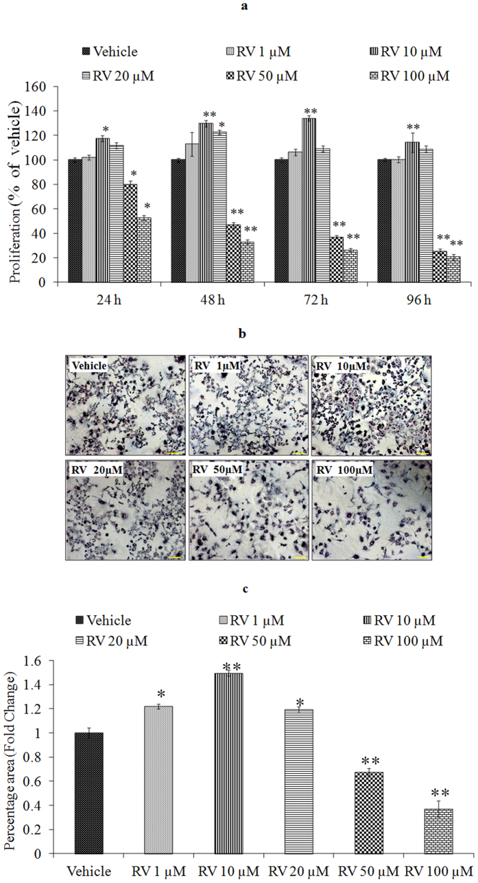
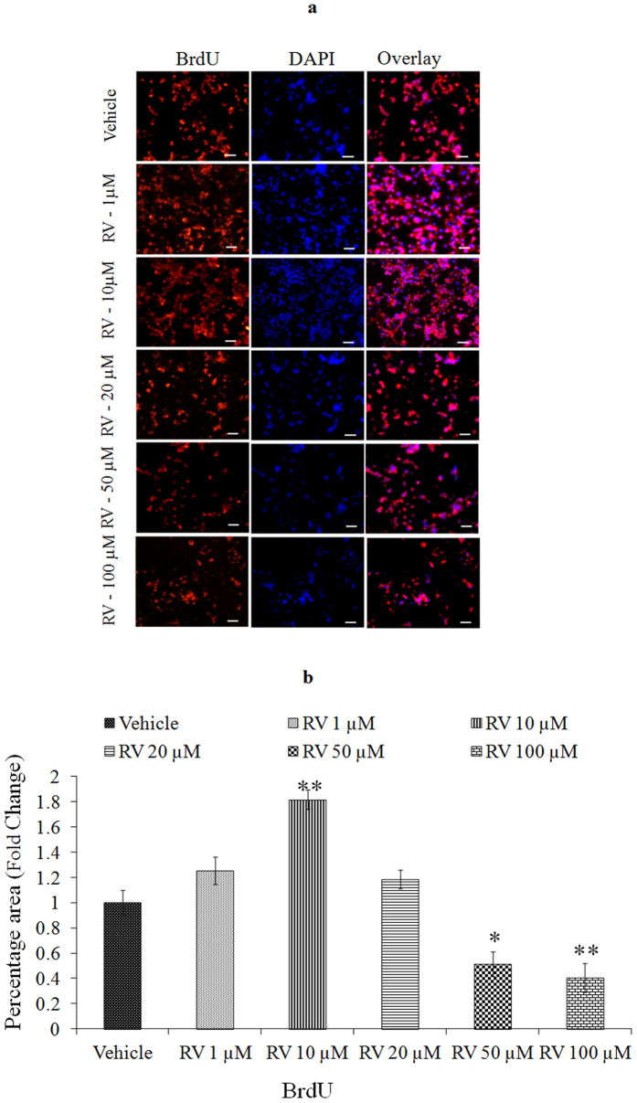
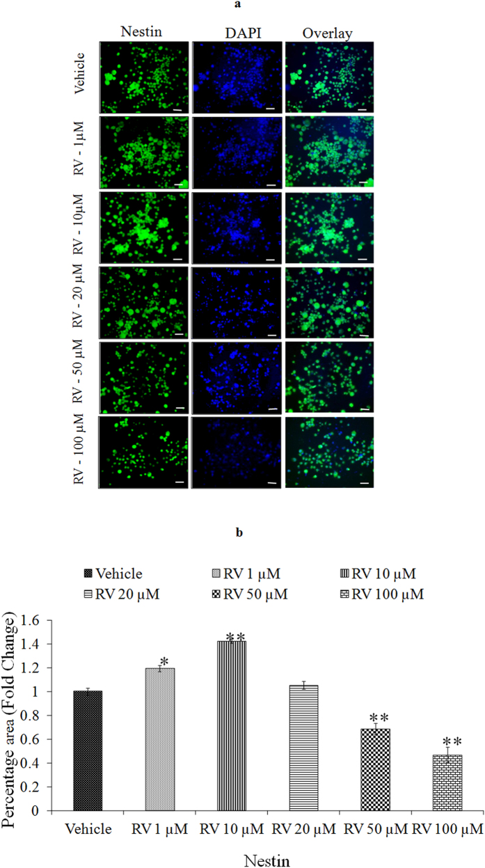
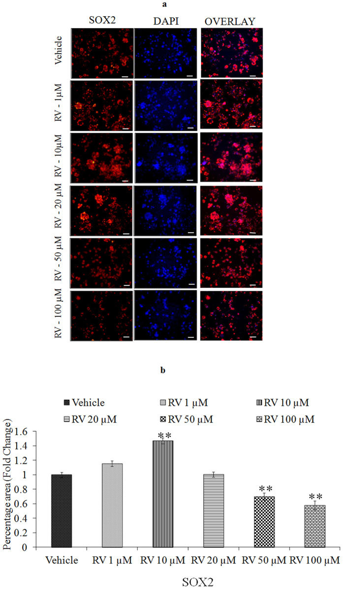
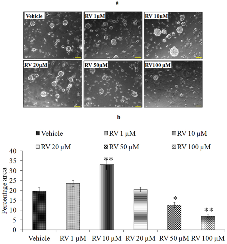
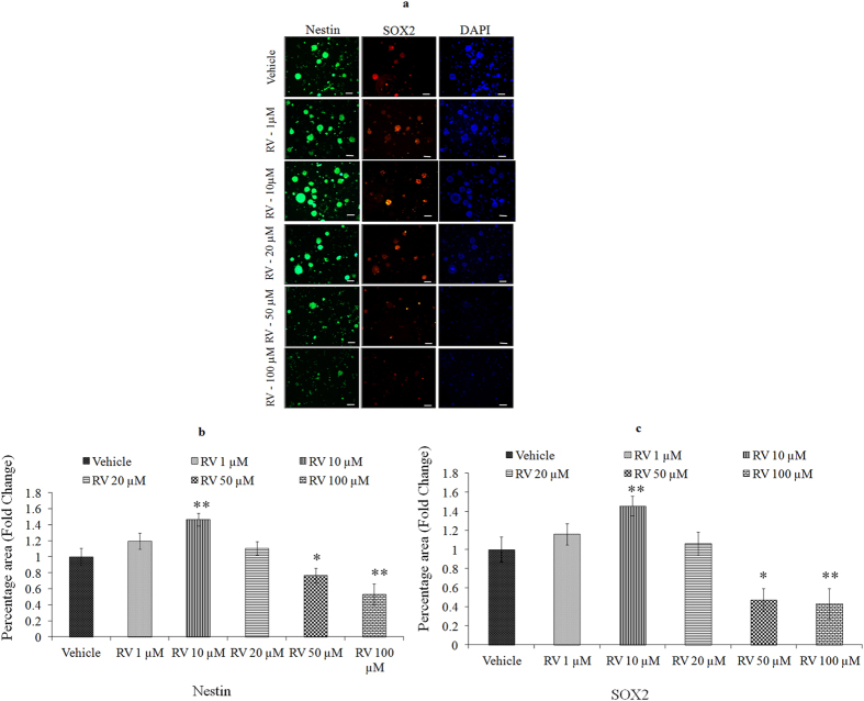
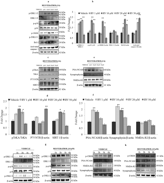
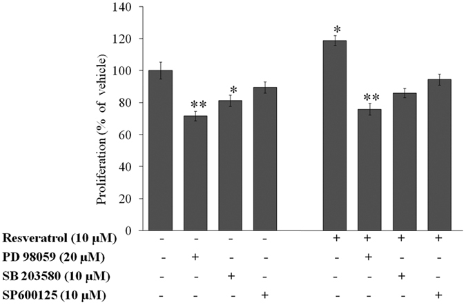
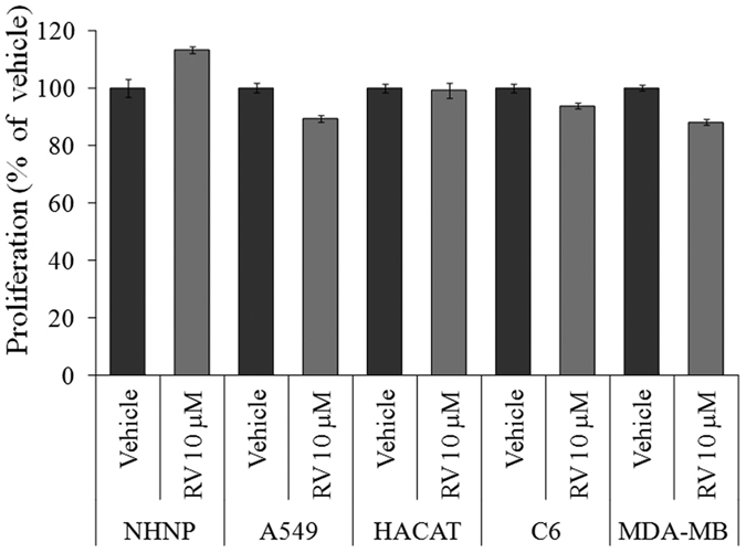
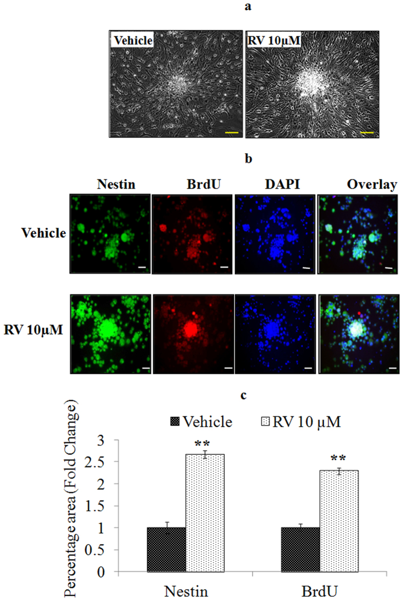
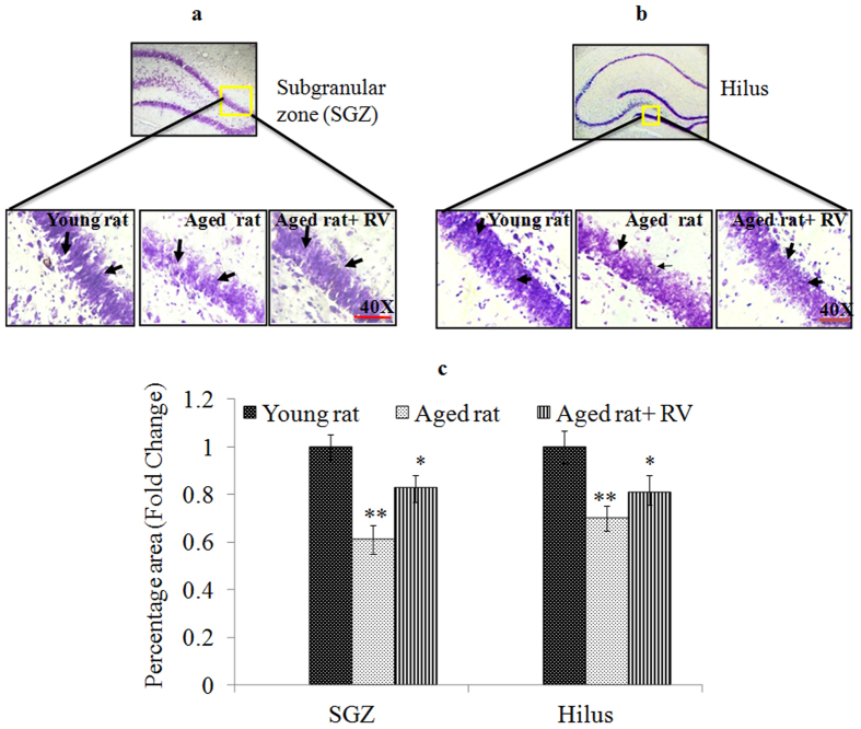
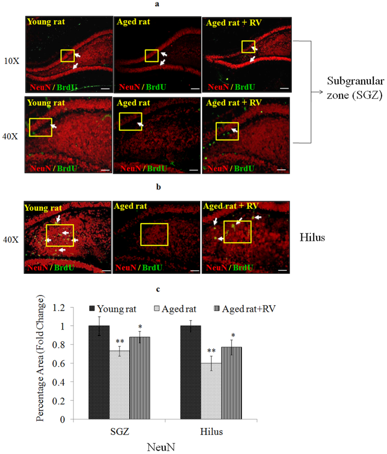
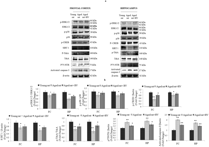
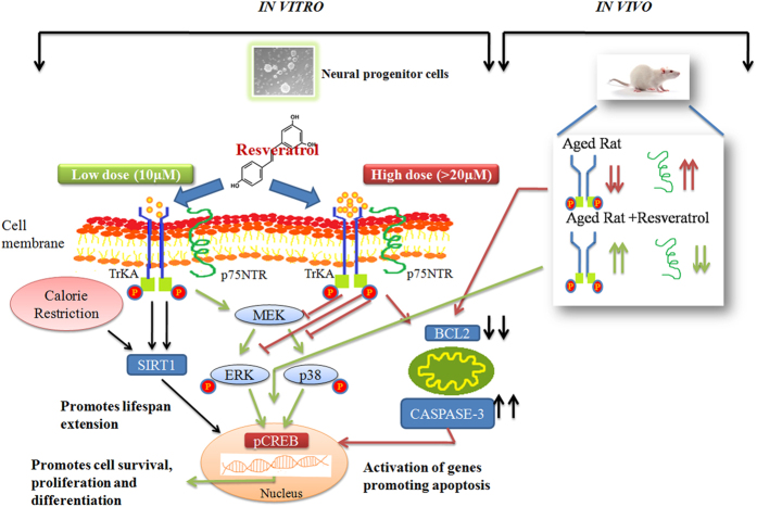
The plethora of literature has supported the potential benefits of Resveratrol (RV) as a life-extending as well as an anticancer compound. However, these two functional discrepancies resulted at different concentration ranges. Likewise, the role of Resveratrol on adult neurogenesis still remains controversial and less understood despite its well documented health benefits. To gather insight into the biological effects of RV on neurogenesis, we evaluated the possible effects of the compound on the proliferation and survival of neural progenitor cells (NPCs) in culture, and in the hippocampus of aged rats. Resveratrol exerted biphasic effects on NPCs; low concentrations (10 μM) stimulated cell proliferation mediated by increased phosphorylation of extracellular signal-regulated kinases (ERKs) and p38 kinases, whereas high concentrations (>20 μM) exhibited inhibitory effects. Administration of Resveratrol (20 mg/kg body weight) to adult rats significantly increased the number of newly generated cells in the hippocampus, with upregulation of p-CREB and SIRT1 proteins implicated in neuronal survival and lifespan extension respectively. We have successfully demonstrated that Resveratrol exhibits dose dependent discrepancies and at a lower concentration can have a positive impact on the proliferation, survival of NPCs and aged rat hippocampal neurogenesis implicating its potential as a candidate for restorative therapies against age related disorders.


Figures

Figure 1
Figure 1. Characterization and proliferation of rNPCs.
(A) Rat brain NPCs isolated from embryonic day-12 (ED-12) rat foetuses showing over 95% viability. (B) Proliferative cells seen as small neurospheres by day 7 in serum free neurobasal medium supplemented with growth factors. (C) The neurospheres attained maturity by day 20. (D,E) Representative micrographs showing expression of both progenitor cell marker-nestin (green) and proliferating cell marker-BrdU (red).
Figure 2
Figure 2. Lower concentrations of Resveratrol stimulates the proliferation of Neural Progenitor cells.
(a) Cell viability assay of NPCs at 24–96 h following the exposure of different concentrations of Resveratrol by MTT assay. (b) Cell viability assay of NPCs by formazon crystal formation at 24 h following the exposure of different concentrations of Resveratrol by MTT assay. Images of formazon crystal were taken after exposure to MTT solution for 4 h. (c) Image quantification was done using ImageJ image analysis software and expressed in fold change. *p < 0.05; **p < 0.01; ***p < 0.001.
Figure 3
Figure 3. Stimulatory effects of lower concentrations of Resveratrol as confirmed by BrdU labelling.
(a) BrdU immunoreactivity (red) in NPCs treated with 20 μM BrdU for 2 h and then exposed to Resveratrol for 24 h. Nuclei were counter stained with DAPI (blue). (b) Image quantification was done using ImageJ image analysis software and expressed in fold change. *p < 0.05; **p < 0.01; ***p < 0.001.
Figure 4
Figure 4. Stimulatory effects of lower concentrations of Resveratrol as confirmed by immunocytochemistry.
(a) Representative microphotographs showing Immunocytochemistry localization of neural progenitor cell marker viz. Nestin (green) in NPCs following the exposure of Resveratrol for 24 h. (b) Image quantification was done using ImageJ image analysis software and expressed in fold change. *p < 0.05; **p < 0.01; ***p < 0.001.
Figure 5
Figure 5. Stimulatory effects of lower concentrations of Resveratrol as confirmed by immunocytochemistry.
(a) Representative microphotographs showing Immunocytochemistry localization of neural progenitor cell marker viz. SOX2 (red) in NPCs following the exposure of Resveratrol for 24 h. (b) Image quantification was done using ImageJ image analysis software and expressed in fold change. *p < 0.05; **p < 0.01; ***p < 0.001.
Figure 6
Figure 6. Lower concentrations of Resveratrol exposure resulted in increase in the number and size of neurospheres.
(a) Embryonic neurospheres were grown in vehicle and different concentration of Resveratrol containing medium for 96 h. Expansion of neurospheres was observed in response to 10 μM of Resveratrol. (b) Image quantification was done using ImageJ image analysis software and expressed in fold change. *p < 0.05; **p < 0.01; ***p < 0.001.
Figure 7
Figure 7. Lower concentrations of Resveratrol increase the expression of proliferation markers.
(a) Represen-tative microphotographs showing Immunocytochemical localization of neural progenitor cell marker viz. Nestin (green) and SOX2 (red) in neurospheres following the exposure of Resveratrol for 96 h. The images were snapped by Nikon DS-Ri1 (12.7megapixel) camera using upright phase contrast florescence microscope (Nikon 80i, Japan) at ×10 × 10 magnification. Data represents mean ± SE of three independent experiments. *p < 0.05; **p < 0.01; ***p < 0.001 (Vehicle vs experimental group). Image quantification (b) Nestin, (c) SOX2 was done using ImageJ image analysis software and expressed in fold change.
Figure 8
Figure 8. Low concentrations of Resveratrol activates ERK1/2 and p38 signalling molecules in NPCs.
(a) Activation of the ERK1/2, p38 and JNK1/2, p-CREB, Bcl-2 and activated caspase-3 on exposure to different concentration of Resveratrol for 1 h. (b) Quantification was done in Gel Documentation System (Alpha Innotech, USA) with the help of AlphaEaseTM FC Stand-Alone V.4.0 software and expressed in fold change. (c) Activation of pTrKA, p75NTR, SIRT 1 on exposure to different concentrations of Resveratrol for 1 h. (d) Quantification was done in Gel Documentation System (Alpha Innotech, USA) with the help of AlphaEaseTM FC Stand-Alone V.4.0 software and expressed in fold change. (e) Activation of downstream genes of p-CREB viz. PSA-NCAM, Synaptophysin and NMDA R2 on exposure of Resveratrol for 1 h. (f) Quantification was done in Gel Documentation System (Alpha Innotech, USA) with the help of AlphaEaseTM FC Stand-Alone V.4.0 software and expressed in fold change. (g) Activation of the ERK1/2, p38 and JNK1/2 signalling molecules phosphorylation on exposure of 10 μM of Resveratrol for different time periods (1–6 h). (h) Activation of p-CREB, PSA-NCAM, Synaptophysin and NMDA R2 proteins on exposure of 10 μM Resveratrol for different time periods (1-6 h)β-actin was used as an internal control to normalize the data. *p < 0.05; **p < 0.01; ***p < 0.001.
Figure 9
Figure 9. Resveratrol induced proliferation of NPCs via activation of ERK1/2 and p38 signalling molecules.
Neural progenitor cells were treated with the indicated kinase inhibitors for 1 h with subsequent treatment with 10 μM Resveratrol for 24 h, and MTT assays were performed. Inhibitors used were: PD98059 (ERK inhibitor), SB203580 (p38 inhibitor), SP600125 (JNK inhibitor). Data represents mean ± SE of three independent experiments. *p < 0.05; **p < 0.01; ***p < 0.001 (Vehicle vs experimental group).
Figure 10
Figure 10. The proliferative action of Resveratrol is selective for neural progenitor cells.
NHNP and the other cell lines (A549, human lung carcinoma; HaCaT, human keratinocytes cell line; C6, rat glioma cells; MDA-MB-231, human breast cell line;) were seeded into 96-well culture plates and cultured for 24 h. The cells were then exposed to 10 μM Resveratrol for 24 h, and cell proliferation was quantified using the MTT assay. Data represents mean ± SE of three independent experiments.
Figure 11
Figure 11. The proliferative action of Resveratrol is mediated through ERK1/2 and p38 signalling molecules activation.
(a) Activation of the ERK1/2, p38 and JNK1/2 signalling molecules phosphorylation on exposure of 10 μM of Resveratrol for 1 h in NHNP and the other cell lines (A549, human lung carcinoma; HACAT, human keratinocytes cell line; C6, rat glioma cells; MDA-MB, human breast cell line;). (b) β-actin was used as an internal control to normalize the data. Quantification of proteins was done in Gel Documentation System (Alpha Innotech, USA) with the help of AlphaEaseTM FC Stand-Alone V.4.0 software and expressed in fold change.
Figure 12
Figure 12. Resveratrol stimulates the proliferation of Normal human neural progenitor (NHNP) cells.
(a) NHNPCs were cultured into 24-well culture plates and treated with Resveratrol (10 μM) for 48 h. Images were taken by Nikon DS-Ri1 (12.7 megapixel) camera at 20 × 10 magnification. (b) Representative photomicrographs showing Immunocytochemistry localization of neural progenitor cell marker viz. Nestin (green) and proliferative marker BrdU (red) in NPCs following the exposure of Resveratrol for 48 h. (c) Image quantification was done using ImageJ image analysis software and expressed in fold change *p < 0.05; **p < 0.01; ***p < 0.001.
Figure 13
Figure 13. Resveratrol stimulates the proliferation in dentate gyrus area of hippocampus in aged rat.
(a) Photomicrographs of SGZ region illustrating Nissl staining in young rat, aged rat and aged rat treated with Resveratrol (20 mg/kg, body weight, p.o.,) for 45 days. (b) Photomicrographs of hilus region illustrating Nissl staining in young rat, aged rat and aged rat treated with Resveratrol (20 mg/kg, body weight, p.o.,) for 45 days. (c) Data represents mean ± SE of three independent experiments. *p < 0.05; **p < 0.01; ***p < 0.001 and is expressed in fold change.
Figure 14
Figure 14. Resveratrol treatment increases the number of BrdU-positive cells in the Dendate gyrus area of hippocampus in aged rat.
(a) Photomicrographs of SGZ region from young, aged and Resveratrol (20 mg/kg, body weight, p.o., for 45 days)-treated aged rats double labeled with fluorescent probes using antibodies against the mature neuron-specific protein (NeuN) and BrdU antibody. BrdU-positive cells (green, proliferation marker); NeuN-positive cells (red, mature neuron maker). (b) Photomicrographs of hilus region from young, aged and Resveratrol (20 mg/kg, body weight, p.o., for 45 days)-treated aged rats double labeled with fluorescent probes using antibodies against the mature neuron-specific protein (NeuN) and BrdU antibody. BrdU-positive cells (green, proliferation marker); NeuN-positive cells (red, mature neuron maker). (c) Data represents mean ± SE of three independent experiments. *p < 0.05; **p < 0.01; ***p < 0.001 and is expressed in fold change.
Figure 15
Figure 15. Resveratrol treatment induces the activation of ERK1/2 and p38 MAP kinases in fontal cortex and hippocampus regions of aged rats.
(a) Protein expression profiling of p-TrkA, p-75NTR, activated caspase-3, MAPK, p-CREB and SIRT1 were studied in fontal cortex and hippocampus regions of young rats, aged rats and Resveratrol (20 mg/kg body weight, p.o., for 45 days) treated aged rats. (b) β-actin was used as internal control to normalize the data. Quantification of proteins was done in Gel Documentation System (Alpha Innotech, USA) with the help of AlphaEaseTM FC StandAlone V.4.0 software and expressed in fold change. *p < 0.05; **p < 0.01; ***p < 0.001.
Figure 16
Figure 16. Resveratrol exerts dose specific biphasic responses.
In vitro- At a low concentration (10 μM) Resveratrol activates the MAPK signalling pathway with a subsequent increase in the expression of p-CREB leading to proliferation, cell survival and differentiation. Resveratrol also independently activates SIRT1 proteins implicated in lifespan extension. However higher doses (>20 μM) inhibit the phosphorylation of the MAPK molecules with a parallel activation of activated caspase-3 inducing the apoptosis pathway. In vivo- The crosstalk between TrkA and p75NTR ligand has a complex role in regulating neural survival and death. Classic signaling modules, such as the MAPK cascade have been identified as downstream cellular events induced by TrkA activation. In aged rats decreased phosphorylation of TrkA is observed with a parallel increase in the expression of the death receptor p75NTR leading to neuronal cell death. Administration of Resveratrol (20 mg/kg body weight) turns the molecular switch around with an increase in phosphorylated levels of TrkA with a subsequent increase in MAPK and p-CREB leading to cell survival and maintenance. This is an original figure made by one of the co-authors Ankita Pandey for publication with this manuscript only.

Similar articles

Cited by

References

    1. Taupin P. & Gage F. H. Adult neurogenesis and neural stem cells of the central nervous system in mammals. Journal of neuroscience research 69, 745–749 (2002). - PubMed
    1. Lee J., Duan W., Long J. M., Ingram D. K. & Mattson M. P. Dietary restriction increases the number of newly generated neural cells, and induces BDNF expression, in the dentate gyrus of rats. Journal of Molecular Neuroscience 15, 99–108 (2000). - PubMed
    1. Lee J., Seroogy K. B. & Mattson M. P. Dietary restriction enhances neurotrophin expression and neurogenesis in the hippocampus of adult mice. Journal of neurochemistry 80, 539–547 (2002). - PubMed
    1. Van Praag H., Christie B. R., Sejnowski T. J. & Gage F. H. Running enhances neurogenesis, learning, and long-term potentiation in mice. Proceedings of the National Academy of Sciences 96, 13427–13431 (1999). - PMC - PubMed
    1. Van Praag H., Kempermann G. & Gage F. H. Running increases cell proliferation and neurogenesis in the adult mouse dentate gyrus. Nature neuroscience 2, 266–270 (1999). - PubMed

Publication types

MeSH terms