. 2016 Aug 12;90(17):7894-901.
doi: 10.1128/JVI.01028-16. Print 2016 Sep 1.

Adeno-associated Virus Vectors Efficiently Transduce Mouse and Rabbit Sensory Neurons Coinfected with Herpes Simplex Virus 1 following Peripheral Inoculation

Affiliations

Adeno-associated Virus Vectors Efficiently Transduce Mouse and Rabbit Sensory Neurons Coinfected with Herpes Simplex Virus 1 following Peripheral Inoculation

Zachary L Watson et al. J Virol. .

Abstract

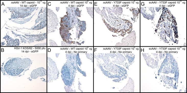
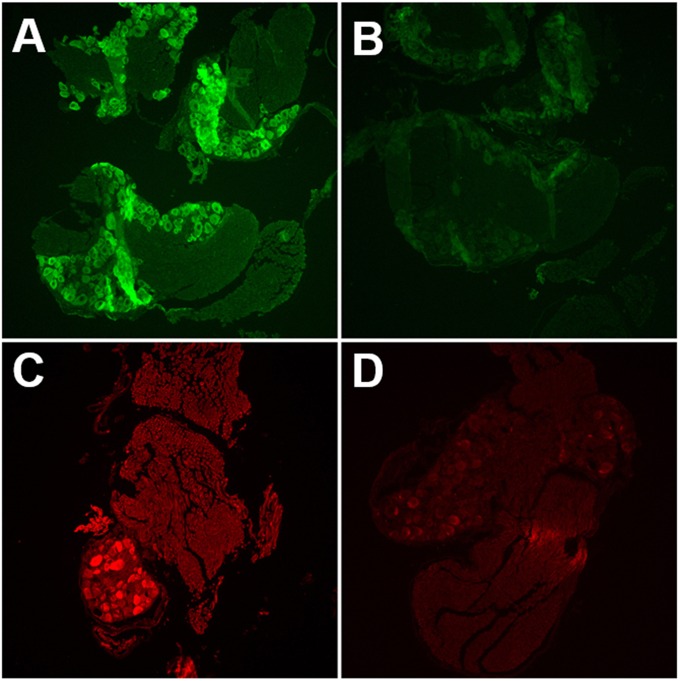
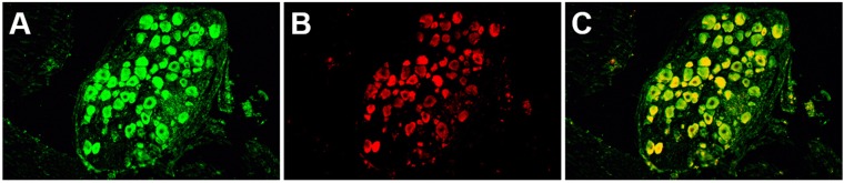
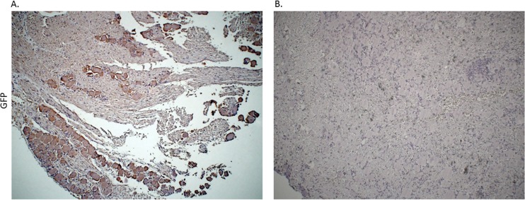
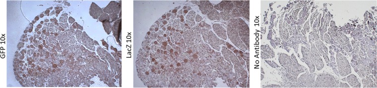
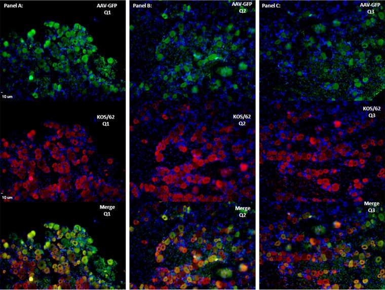
Following infection of epithelial tissues, herpes simplex virus 1 (HSV-1) virions travel via axonal transport to sensory ganglia and establish a lifelong latent infection within neurons. Recent studies have revealed that, following intraganglionic or intrathecal injection, recombinant adeno-associated virus (rAAV) vectors can also infect sensory neurons and are capable of stable, long-term transgene expression. We sought to determine if application of rAAV to peripheral nerve termini at the epithelial surface would allow rAAV to traffic to sensory ganglia in a manner similar to that seen with HSV. We hypothesized that footpad or ocular inoculation with rAAV8 would result in transduction of dorsal root ganglia (DRG) or trigeminal ganglia (TG), respectively. To test this, we inoculated the footpads of mice with various amounts of rAAV as well as rAAV capsid mutants. We demonstrated that this method of inoculation can achieve a transduction rate of >90% of the sensory neurons in the DRG that innervate the footpad. Similarly, we showed that corneal inoculation with rAAV vectors in the rabbit efficiently transduced >70% of the TG neurons in the optic tract. Finally, we demonstrated that coinfection of mouse footpads or rabbit eyes with rAAV vectors and HSV-1 resulted in colocalization in nearly all of the HSV-1-positive neurons. These results suggest that rAAV is a useful tool for the study of HSV-1 infection and may provide a means to deliver therapeutic cargos for the treatment of HSV infections or of dysfunctions of sensory ganglia.

Importance: Adeno-associated virus (AAV) has been shown to transduce dorsal root ganglion sensory neurons following direct intraganglionic sciatic nerve injection and intraperitoneal and intravenous injection as well as intrathecal injection. We sought to determine if rAAV vectors would be delivered to the same sensory neurons that herpes simplex virus (HSV-1) infects when applied peripherally at an epithelial surface that had been treated to expose the underlying sensory nerve termini. For this study, we chose two well-established HSV-1 infection models: mouse footpad infection and rabbit ocular infection. The results presented here provide the first description of AAV vectors transducing neurons following delivery at the skin/epithelium/eye. The ability of AAV to cotransduce HSV-1-infected neurons in both the mouse and the rabbit provides an opportunity to experimentally explore and disrupt host and viral proteins that are integral to the establishment of HSV-1 latency, to the maintenance of latency, and to reactivation from latency in vivo.


Figures

FIG 1
FIG 1
Following footpad inoculation, rAAV8 vectors transduce nearly 100% of sensory neurons in mouse DRG. Vectors with modified capsids are at least 10-fold more efficient than wild-type vectors. To analyze the immunohistochemistry of infected mouse DRG, mice were infected with rAAV or KOS/62 at the given dose and tissues were harvested as shown. Anti-GFP primary antibody was incubated with sections A, B, C, E, and G to detect the presence of GFP. No primary antibody was incubated with sections D, F, and H. All sections were stained by HRP enzymatic cleavage of DAB substrate (brown) for 8 min. Hematoxylin (blue) was used as a counterstain.
FIG 2
FIG 2
Single-color immunofluorescence detects both rAAV8 and HSV-1 in mouse DRG. (A and B) Mice were infected with ssAAV-GFP only. DRG were harvested 14 dpi. Tissue sections were incubated with anti-GFP primary antibody (A) or with no primary antibody (B). Primary antibody was detected by the use of FITC-labeled secondary antibody. (C and D) Mice were infected with KOS/62 only. DRG were harvested 4 dpi. Tissue sections were incubated with anti-β-Gal primary antibody (C) or with no primary antibody (D). Primary antibody was detected by Texas Red-labeled secondary antibody.
FIG 3
FIG 3
Dual-color immunofluorescence demonstrates colocalization of rAAV8 and HSV-1 within a subset of sensory neurons in mouse DRG. Mice were infected with both 1010 particles of scAAV-GFP-WT and 5,000 PFU of KOS/62. DRG were harvested 4 dpi. Tissue sections were incubated with both anti-GFP and anti-β-Gal primary antibodies. (A) Green channel image. Anti-GFP primary antibody was detected by FITC-labeled secondary antibody. Green cells were transduced by AAV. (B) Red channel image. Anti-β-Gal primary antibody was detected by Texas Red-labeled secondary antibody. Red cells were infected by HSV-1. (C) Merged image representing the red and green images. Yellow indicates colocalization of GFP and β-Gal expression from cells coinfected with both AAV and HSV-1.
FIG 4
FIG 4
Immunohistochemistry analysis of rabbit TGs following corneal delivery of AAV8-GFP capsid mutant confirmed that AAV8 efficiently transduces neurons through a corneal route of delivery. Immunohistochemistry was done using rabbit TG following corneal inoculation with ssAAV8-GFP-Y733. All TG were harvested on postinoculation day 16. (A) Primary antibody incubation was done using mouse anti-GFP (Abcam), followed by incubation with a biotinylated HRP secondary antibody (Vector Laboratories). (B) The control slide was prepared from the TG of naive rabbit without AAV treatment and was subjected to both primary and secondary antibody incubation.
FIG 5
FIG 5
Immunohistochemistry using rabbit TGs following corneal delivery of ssAAV8-GFP-Y733 and a KOS/62 demonstrates colocalization of both the vector and HSV-1 in sensory neurons. Serial sections of TG from rabbits inoculated with ssAAV8-GFP-Y733 and KOS/62 were used to demonstrate that the vector and HSV-1 colocalize in neurons. Serial sections of <5 μm are presented at magnifications of ×10. IHC to detect the presence of KOS/62 was done using a mouse anti-β-galactosidase primary antibody (Abcam), followed by a biotinylated HRP secondary antibody (Vector Laboratories). To detect AAV8-GFP in neurons, sections were incubated with mouse anti-GFP antibody (Abcam), followed by incubation with a biotinylated HRP secondary antibody (Vector Laboratories). IHC was done to detect either HSV-1 or AAV8 using serial sections to demonstrate that neurons harbor both the vector and the virus. Control slides were prepared from coinfected rabbit TG, subjected to IHC with no primary antibody.
FIG 6
FIG 6
Immunofluorescence results showed long-term expression and colocalization of AAV8-GFP and KOS/62 in coinfected rabbit TG. IF analysis was performed to demonstrate neuronal colocalization of vector and virus. Sections were incubated with either mouse anti-GFP or mouse anti-β-galactosidase primary antibodies. Alexa Fluor 548 (Invitrogen) or goat anti-mouse Alexa Fluor 488 (Invitrogen) was used as the secondary antibody for immunofluorescent visualization using a Leica deconvolution microscope with Slidebook 5.0. The TG section on the slide was divided into three equal sections (Q1 to Q3 [A to C]) to represent the colocalization of virus and vector over the entire TG cross-section. DAPI (4′,6-diamidino-2-phenylindole) staining (in blue) represents nuclei of satellite cells present in the sections. All images are shown in ×10 magnification.

Similar articles

Cited by

References

    1. Margolis TP, Dawson CR, LaVail JH. 1992. Herpes simplex viral infection of the mouse trigeminal ganglion. Immunohistochemical analysis of cell populations. Invest Ophthalmol Vis Sci 33:259–267. - PubMed
    1. Yang L, Voytek CC, Margolis TP. 2000. Immunohistochemical analysis of primary sensory neurons latently infected with herpes simplex virus type 1. J Virol 74:209–217. doi:10.1128/JVI.74.1.209-217.2000. - DOI - PMC - PubMed
    1. Russell DW, Miller AD, Alexander IE. 1994. Adeno-associated virus vectors preferentially transduce cells in S phase. Proc Natl Acad Sci U S A 91:8915–8919. doi:10.1073/pnas.91.19.8915. - DOI - PMC - PubMed
    1. Lovric J, Mano M, Zentilin L, Eulalio A, Zacchigna S, Giacca M. 2012. Terminal differentiation of cardiac and skeletal myocytes induces permissivity to AAV transduction by relieving inhibition imposed by DNA damage response proteins. Mol Ther 20:2087–2097. doi:10.1038/mt.2012.144. - DOI - PMC - PubMed
    1. Ferrari FK, Samulski T, Shenk T, Samulski RJ. 1996. Second-strand synthesis is a rate-limiting step for efficient transduction by recombinant adeno-associated virus vectors. J Virol 70:3227–3234. - PMC - PubMed

Publication types