. 2016 Aug 12;90(17):7864-79.
doi: 10.1128/JVI.00869-16. Print 2016 Sep 1.

Opposing Roles of Double-Stranded RNA Effector Pathways and Viral Defense Proteins Revealed with CRISPR-Cas9 Knockout Cell Lines and Vaccinia Virus Mutants

Affiliations

Opposing Roles of Double-Stranded RNA Effector Pathways and Viral Defense Proteins Revealed with CRISPR-Cas9 Knockout Cell Lines and Vaccinia Virus Mutants

Ruikang Liu et al. J Virol. .

Abstract

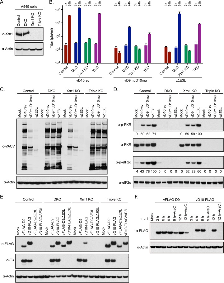
Vaccinia virus (VACV) decapping enzymes and cellular exoribonuclease Xrn1 catalyze successive steps in mRNA degradation and prevent double-stranded RNA (dsRNA) accumulation, whereas the viral E3 protein can bind dsRNA. We showed that dsRNA and E3 colocalized within cytoplasmic viral factories in cells infected with a decapping enzyme mutant as well as with wild-type VACV and that they coprecipitated with antibody. An E3 deletion mutant induced protein kinase R (PKR) and eukaryotic translation initiation factor alpha (eIF2α) phosphorylation earlier and more strongly than a decapping enzyme mutant even though less dsRNA was made, leading to more profound effects on viral gene expression. Human HAP1 and A549 cells were genetically modified by clustered regularly interspaced short palindromic repeat-Cas9 (CRISPR-Cas9) to determine whether the same pathways restrict E3 and decapping mutants. The E3 mutant replicated in PKR knockout (KO) HAP1 cells in which RNase L is intrinsically inactive but only with a double knockout (DKO) of PKR and RNase L in A549 cells, indicating that both pathways decreased replication equivalently and that no additional dsRNA pathway was crucial. In contrast, replication of the decapping enzyme mutant increased significantly (though less than that of wild-type virus) in DKO A549 cells but not in DKO HAP1 cells where a smaller increase in viral protein synthesis occurred. Xrn1 KO A549 cells were viable but nonpermissive for VACV; however, wild-type and mutant viruses replicated in triple-KO cells in which RNase L and PKR were also inactivated. Since KO of PKR and RNase L was sufficient to enable VACV replication in the absence of E3 or Xrn1, the poor replication of the decapping mutant, particularly in HAP1 DKO, cells indicated additional translational defects.

Importance: Viruses have evolved ways of preventing or counteracting the cascade of antiviral responses that double-stranded RNA (dsRNA) triggers in host cells. We showed that the dsRNA produced in excess in cells infected with a vaccinia virus (VACV) decapping enzyme mutant and by wild-type virus colocalized with the viral E3 protein in cytoplasmic viral factories. Novel human cell lines defective in either or both protein kinase R and RNase L dsRNA effector pathways and/or the cellular 5' exonuclease Xrn1 were prepared by CRISPR-Cas9 gene editing. Inactivation of both pathways was necessary and sufficient to allow full replication of the E3 mutant and reverse the defect cause by inactivation of Xrn1, whereas the decapping enzyme mutant still exhibited defects in gene expression. The study provided new insights into functions of the VACV proteins, and the well-characterized panel of CRISPR-Cas9-modified human cell lines should have broad applicability for studying innate dsRNA pathways.


Figures

FIG 1
FIG 1
Colocalization of dsRNA and E3 protein in infected cells. (A) A549 cells on coverslips were mock infected or infected with 5 PFU per cell of vD10rev, vD9muD10mu, or vΔE3L. After 13 h, the cells were fixed, permeabilized, and stained for dsRNA with J2 MAb (green) and for E3 with E3 MAb (red), followed by isotype-specific IgG2A and IgG3 fluorescent secondary antibodies, respectively, and DAPI. The cells were imaged by confocal microscopy. Arrows point to virus factories. The factory region within the boxed area was enlarged and is shown in the row below. Scale bar, 10 μm. (B) A549 cells were infected as described for panel A, stained with J2 MAb and fluorescent secondary antibody, and analyzed by flow cytometry. (C) The experiment is the same as that described for panel A except that cells were mock infected or infected with vD10rev-E3-GFP or vD9muD10mu-E3-GFP and stained with J2 MAb to detect dsRNA (red) followed by secondary fluorescent antibodies. E3 was visualized by GFP fluorescence. The factory region in the boxed area was enlarged and is shown in the row below. (D) A549 cells were infected with vD9muD10mu-E3-GFP, and lysates were analyzed by Western blotting with antibody to GFP and RIG-I or immunoprecipitated (IP) with J2 MAb or nonspecific IgG and then analyzed by Western blotting.
FIG 2
FIG 2
Replication of decapping enzyme mutant and E3 deletion virus in HAP1 and A549 KO cells. (A) Absence of RNase L and PKR proteins in HAP1 KO cells. Cell lysates from HAP1 control, RNase L KO, PKR KO, and DKO cells were analyzed by Western blotting using mouse polyclonal antibody to RNase L and rabbit MAb to PKR. Antibody to actin was used as a loading control. (B) One-step virus replication. HAP1 control, RNase L KO, PKR KO, and DKO cell monolayers in 12-well plates were infected in triplicate with 5 PFU/cell of purified vD10rev, vD9muD10mu, or vΔE3L and harvested at 3 and 24 h. Virus titers were determined by plaque assay in BHK-21 cells. The 3-h titers represent input virus. (C and D) Procedures were the same as those described for panels A and B except that A549 control and KO cells were used. Each bar represents the standard deviation determined from three replicate infections.
FIG 3
FIG 3
Transmission electron microscopy. DKO A549 (upper two rows) and HAP1 cells (lower two rows) were infected with 5 PFU per cell of vD10rev or vD9muD10mu and harvested after 13 h. The cells were fixed, cut into thin sections, and examined by transmission electron microcopy. IV, immature virion; MV, mature virion; DV, aberrant dense virion.
FIG 4
FIG 4
Viral gene expression in HAP1 KO cells. (A to D) Western blotting. Control, RNase L KO, PKR KO, and DKO HAP1 cells were infected with 5 PFU/cell of purified vD10rev, vD9muD10mu, or vΔE3L and harvested at the indicated times (shown in hours). The lysates were analyzed by Western blotting using antibodies to VACV (A), the E3 early protein (B), the D13 intermediate protein (C), and the A3 late protein (D). Antibody to actin served as a loading control (not shown). One set of cells was infected in the presence of AraC to inhibit viral DNA replication and confirm the stage of expression of the viral proteins. (E) Northern blotting. HAP1 control, RNase L KO, PKR KO, and DKO cells were mock infected or infected with 5 PFU/cell of purified vD10rev, vD9muD10mu, or vΔE3L virus. At 13 h after infection, the cells were harvested, and the total RNAs were isolated and resolved on glyoxal gels. rRNAs were stained with ethidium bromide and detected by UV fluorescence; reverse images are shown. The RNAs were transferred to a nylon membrane, incubated with the digoxigenin-labeled probes to the viral early C11 mRNA, viral late F17 mRNA, and cellular GAPDH mRNA, detected with alkaline phosphatase-conjugated antibody to digoxigenin, and visualized with the chemiluminescence substrate and X-ray film. p.i., postinfection; Ab, antibody.
FIG 5
FIG 5
Innate immune responses in HAP1 KO cells. (A) PKR. Control, RNase L KO, PKR KO, and DKO HAP1 cells were infected with 5 PFU/cell of purified vD10rev, vD9muD10mu, or vΔE3L in the absence or presence of AraC and harvested at the indicated hour after infection. The lysates were analyzed by SDS-PAGE and Western blotting using antibodies to phosphorylated PKR (α-p-PKR). Densities shown below the images were determined with ImageJ. (B) eIF2α. A Western blot prepared as described for panel A was probed with antibodies to phosphorylated eIF2α (α-p-eIF2α).
FIG 6
FIG 6
Viral gene expression and innate immune response in A549 KO cells. (A) Viral protein synthesis. A549 control, RNase L KO, PKR KO, and DKO cells were mock infected or infected with 5 PFU/cell of purified vD10rev, vD9muD10mu, or vΔE3L. At 13 h after infection, the cell lysates were analyzed by Western blotting using rabbit polyclonal antibody to VACV proteins and actin. (B) E3 synthesis. Control and KO A549 cells were mock infected or infected with vD10rev, vD9muD10mu, or vΔE3L in the absence or presence of AraC. The cells were harvested at 2, 4, 8, and 12 h after infection and analyzed by Western blotting with a MAb to E3. (C) rRNA and mRNA. Total RNA was isolated from cells infected as described for panel A and resolved on glyoxal gels. The RNAs were stained with ethidium bromide and detected by UV fluorescence; reverse images are shown. RNAs were transferred from the glyoxal gel to a nylon membrane, incubated with the digoxigenin-labeled probes to the viral late F17 mRNA or cellular GAPDH mRNA, detected with alkaline phosphatase-conjugated antibody to digoxigenin, and visualized with a chemiluminescence substrate on X-ray films. (D) IRF3. Lysates from cells infected as described for panel A were analyzed by Western blotting with antibodies to phosphorylated IRF3, IRF3 protein, and actin. Densities shown below the images were determined with ImageJ.
FIG 7
FIG 7
Synthesis of dsRNA in Xrn1 KO cells. (A) Control and Xrn1 KO A549 cells were mock infected or infected with 5 PFU per cell of purified vD10rev, vD9muD10mu, or vΔE3L, harvested after 13 h, and stained with J2 MAb and a secondary fluorescent antibody. Fluorescent cells were analyzed by flow cytometry. (B) A549 Xrn1 KO cells were infected as described for panel A, stained with J2 MAb, secondary fluorescent antibody, and DAPI, and examined by confocal microscopy. Scale bar, 10 µm.
FIG 8
FIG 8
Replication of VACV in Xrn1 and Xrn1+PKR+RNase L TKO A549 cells. (A) Analysis of KO cells. A549 cells and A549 PKR+RNase L DKO cells were used to make Xrn1 KO and Xrn1+PKR+RNase L triple-KO (TKO) cells. Western blotting with Xrn1 and actin antibodies confirmed inactivation of the Xrn1 gene. (B) Virus replication. Control A549, DKO, Xrn1 KO, and TKO cells were infected with 5 PFU/cell of purified vD10rev, vD9muD10mu, and vΔE3L. Cells were lysed at 3 and 24 h to determine the virus input and yield, respectively, by plaque assay on BHK-21 cells. (C) Viral protein synthesis. A549 control, DKO, two independent Xrn1 KO, and TKO cells were infected with 5 PFU per cell of vD10rev, vD9muD10mu, or vΔE3L and harvested after 13 h. Lysates were analyzed by Western blotting using antibodies to VACV and actin. (D) PKR and eIF2α phosphorylation. Cells were infected as described for panel C, and Western blots were probed with antibody to phosphorylated PKR, PKR protein, phosphorylated eIF2α, and eIF2α protein. Densities shown below the images were determined with ImageJ. (E) D9 and D10 synthesis. A549 control, DKO, Xrn1 KO, and TKO cells were mock infected or infected with 5 PFU/cell of vFLAG-D9, vD10-FLAG, vFLAG-D9ΔE3L, or vD10-FLAGΔE3L. At 13 h after infection, the cells were harvested, and lysates were analyzed by Western blotting using antibodies to FLAG and E3. (F) Time course of D9 and D10 synthesis. A549 cells were mock infected or infected with vFLAG-D9 or vD10-FLAG in the absence of presence of AraC. At the indicated hours after infection, the cells were harvested, and lysates were analyzed by Western blotting with antibody to the FLAG epitope or actin.

Similar articles

Cited by

References

    1. Silverman RH. 2007. Viral encounters with 2 ′,5′-oligoadenylate synthetase and RNase L during the interferon antiviral response. J Virol 81:12720–12729. doi: 10.1128/JVI.01471-07. - DOI - PMC - PubMed
    1. DeWitte-Orr SJ, Mossman KL. 2010. dsRNA and the innate antiviral immune response. Future Virol 5:325–341. doi: 10.2217/fvl.10.18. - DOI
    1. Drappier M, Michiels T. 2015. Inhibition of the OAS/RNase L pathway by viruses. Curr Opin Virol 15:19–26. doi: 10.1016/j.coviro.2015.07.002. - DOI - PMC - PubMed
    1. Duesberg PH, Colby C. 1969. On the biosynthesis and structure of double-stranded RNA in vaccinia virus-infected cells. Proc Natl Acad Sci U S A 64:396–403. doi: 10.1073/pnas.64.1.396. - DOI - PMC - PubMed
    1. Liu SW, Katsafanas GC, Liu R, Wyatt LS, Moss B. 2015. Poxvirus decapping enzymes enhance virulence by preventing the accumulation of dsRNA and the induction of innate antiviral responses. Cell Host Microbe 17:320–331. doi: 10.1016/j.chom.2015.02.002. - DOI - PMC - PubMed

Publication types

MeSH terms