

Cell-Free versus Cell-to-Cell Infection by Human Immunodeficiency Virus Type 1 and Human T-Lymphotropic Virus Type 1: Exploring the Link among Viral Source, Viral Trafficking, and Viral Replication
- PMID: 27334587
- PMCID: PMC4988172
- DOI: 10.1128/JVI.00407-16
Cell-Free versus Cell-to-Cell Infection by Human Immunodeficiency Virus Type 1 and Human T-Lymphotropic Virus Type 1: Exploring the Link among Viral Source, Viral Trafficking, and Viral Replication
Abstract
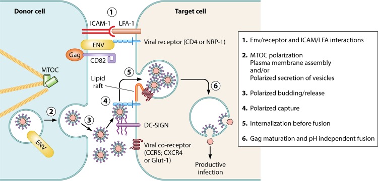
Human immunodeficiency virus type 1 (HIV-1) and human T-lymphotropic virus type 1 (HTLV-1) are complex retroviruses mainly infecting CD4(+) T lymphocytes. In addition, antigen-presenting cells such as dendritic cells (DCs) are targeted in vivo by both viruses, although to a lesser extent. Interaction of HIV-1 with DCs plays a key role in viral dissemination from the mucosa to CD4(+) T lymphocytes present in lymphoid organs. While similar mechanisms may occur for HTLV-1 as well, most HTLV-1 data were obtained from T-cell studies, and little is known regarding the trafficking of this virus in DCs. We first compared the efficiency of cell-free versus cell-associated viral sources of both retroviruses at infecting DCs. We showed that both HIV-1 and HTLV-1 cell-free particles are poorly efficient at productively infecting DCs, except when DC-SIGN has been engaged. Furthermore, while SAMHD-1 accounts for restriction of cell-free HIV-1 infection, it is not involved in HTLV-1 restriction. In addition, cell-free viruses lead mainly to a nonproductive DC infection, leading to trans-infection of T-cells, a process important for HIV-1 spread but not for that of HTLV-1. Finally, we show that T-DC cell-to-cell transfer implies viral trafficking in vesicles that may both increase productive infection of DCs ("cis-infection") and allow viral escape from immune surveillance. Altogether, these observations allowed us to draw a model of HTLV-1 and HIV-1 trafficking in DCs.
Copyright © 2016, American Society for Microbiology. All Rights Reserved.
Figures


References
-
- UNAIDS. 2013. AIDS by the numbers. www.unaids.org UNAIDS, Washington, DC.
-
- Bruhn R, Mahieux R, Murphy EL. Human lymphotropic viruses: HTLV-1 and HTLV-2. In clinical virology, 4th ed, in press ASM Press, Washington, DC.
-
- Uchiyama T, Yodoi J, Sagawa K, Takatsuki K, Uchino H. 1977. Adult T-cell leukemia: clinical and hematologic features of 16 cases. Blood 50:481–492. - PubMed
-
- Gessain A, Barin F, Vernant J, Gout O, Maurs L, Calender A, de Thé G. 1985. Antibodies to human T-lymphotropic virus type-I in patients with tropical spastic paraparesis. Lancet ii:407–410. - PubMed
Publication types
MeSH terms
LinkOut - more resources
Full Text Sources
Other Literature Sources
Research Materials
Miscellaneous
