Review
. 2016 Jun 22:13:107.
doi: 10.1186/s12985-016-0561-z.

Biological function of Foot-and-mouth disease virus non-structural proteins and non-coding elements

Affiliations
Review

Biological function of Foot-and-mouth disease virus non-structural proteins and non-coding elements

Yuan Gao et al. Virol J. .

Abstract

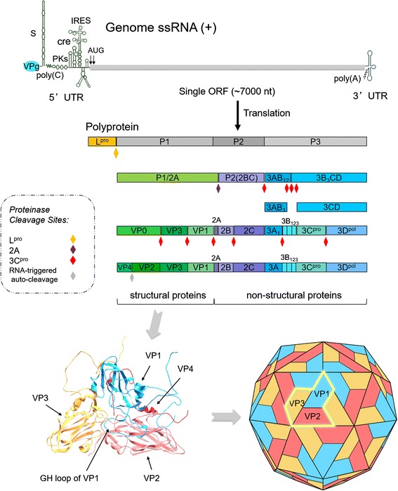
Foot-and-mouth disease virus (FMDV) represses host translation machinery, blocks protein secretion, and cleaves cellular proteins associated with signal transduction and the innate immune response to infection. Non-structural proteins (NSPs) and non-coding elements (NCEs) of FMDV play a critical role in these biological processes. The FMDV virion consists of capsid and nucleic acid. The virus genome is a positive single stranded RNA and encodes a single long open reading frame (ORF) flanked by a long structured 5'-untranslated region (5'-UTR) and a short 3'-UTR. The ORF is translated into a polypeptide chain and processed into four structural proteins (VP1, VP2, VP3, and VP4), 10 NSPs (L(pro), 2A, 2B, 2C, 3A, 3B1-3, 3C(pro), and 3D(pol)), and some cleavage intermediates. In the past decade, an increasing number of studies have begun to focus on the molecular pathogenesis of FMDV NSPs and NCEs. This review collected recent research progress on the biological functions of these NSPs and NCEs on the replication and host cellular regulation of FMDV to understand the molecular mechanism of host-FMDV interactions and provide perspectives for antiviral strategy and development of novel vaccines.


Figures

Fig. 1
Fig. 1
Schematic diagram of FMDV genome, processing of viral polypeptide and conformations of the structural proteins. FMDV genome RNA contains a single open reading frame (ORF) of about 7 kb with two alternative initiation sites. The ORF is flanked by a long 5ʹ-untranslated region (5ʹ-UTR) and a short 3ʹ-UTR. 3B (VPg) is covalently bound to its 5ʹ end. The ORF region is generally divided into four functional areas (L, P1, P2 and P3) due to the different functions of mature polypeptides. ORF-encoded polyprotein is processed into four products, Lpro, P1-2A, 2BC and P3 by Lpro, 2A and 3Cpro. The precursors P1-2A, 2BC and P3 are further processed into mature viral proteins and some cleavage intermediates with relative stability, such as VP0 or 1AB, 3ABC, 3BCD, 3AB, and 3CD by 3Cpro. Structural proteins form the biological protomer and viral capsid
Fig. 2
Fig. 2
Life cycle of FMDV in host cells. NSPs, non-structural proteins. HS, heparan sulfate. Green line, viral positive-strand (+) RNA. Orange line, viral negative-strand (-) RNA
Fig. 3
Fig. 3
FMDV 5ʹ-UTR provides structural basis for initiation of viral protein translation. a Schematic representation of the structural elements within the FMDV 5ʹ-UTR. The 5ʹ-UTR is highly structured and comprises S-fragment, poly(C) tract, 3B-uridylylation site (bus) or cis-acting replication element (cre), pseudoknots (PKs), and internal ribosome entry site (IRES). The organization of the IRES element in different domains (I, II, III, IV, and V) is indicated. Domain I (the first 20 nt of the IRES) belongs to the right arm of bus. The conserved AAACA, GNRA, RAAA motifs, C-rich loop and polypyrimidine tracts are also indicated. The two different initiator codons AUG1 and AUG2 are separated by 84 nt. Note that the stems indicated are not perfectly base-paired. b Intact eIF4G is required in cap-dependent protein synthesis of cellular mRNA. The initiation factors are required for the assembly of 48S initiation complex on a capped cellular mRNA. c Involvement of translation initiation factors in IRES-dependent protein synthesis. The FMDV IRES elements are indicated. The factors shown are eIF4E (4E), eIF4G (4G), eIF4A (4A), and eIF2 (2) (as part of the ternary complex eIF2/GTP/met-tRNA), along with small ribosomal subunit (40S)
Fig. 4
Fig. 4
Known functions of FMDV nonstructural proteins on cellular regulation. Purple circles: FMDV nonstructural proteins. Red arrow: activation of downstream gene expression. Orange line with vertical stub: inhibition of downstream proteins. kB, a canonical kB element response to Nuclear factor kB (NF-kB). ISRE, IFN-sensitive response element. Caspases, represent a class of genes inducing apoptosis and inflammation

References

    1. Knowles NJ, Samuel AR. Molecular epidemiology of foot-and-mouth disease virus. Virus Res. 2003;91:65–80. doi: 10.1016/S0168-1702(02)00260-5. - DOI - PubMed
    1. Haydon DT, Samuel AR, Knowles NJ. The generation and persistence of genetic variation in foot-and-mouth disease virus. Prev Vet Med. 2001;51:111–24. doi: 10.1016/S0167-5877(01)00210-0. - DOI - PubMed
    1. Domingo E, Ruiz-Jarabo CM, Sierra S, Arias A, Pariente N, Baranowski E, Escarmis C. Emergence and selection of RNA virus variants: memory and extinction. Virus Res. 2002;82:39–44. doi: 10.1016/S0168-1702(01)00385-9. - DOI - PubMed
    1. Domingo E, Pariente N, Airaksinen A, Gonzalez-Lopez C, Sierra S, Herrera M, Grande-Perez A, Lowenstein PR, Manrubia SC, Lazaro E, Escarmis C. Foot-and-mouth disease virus evolution: exploring pathways towards virus extinction. Curr Top Microbiol Immunol. 2005;288:149–73. - PMC - PubMed
    1. Klein J. Understanding the molecular epidemiology of foot-and-mouth-disease virus. Infect Genet Evol. 2009;9:153–61. doi: 10.1016/j.meegid.2008.11.005. - DOI - PMC - PubMed

Publication types

MeSH terms

Substances

LinkOut - more resources