. 2016 Jun;145(6):757-68.
doi: 10.1093/ajcp/aqw075.

Enhanced Islet Cell Nucleomegaly Defines Diffuse Congenital Hyperinsulinism in Infancy but Not Other Forms of the Disease

Affiliations

Enhanced Islet Cell Nucleomegaly Defines Diffuse Congenital Hyperinsulinism in Infancy but Not Other Forms of the Disease

Bing Han et al. Am J Clin Pathol. 2016 Jun.

Abstract

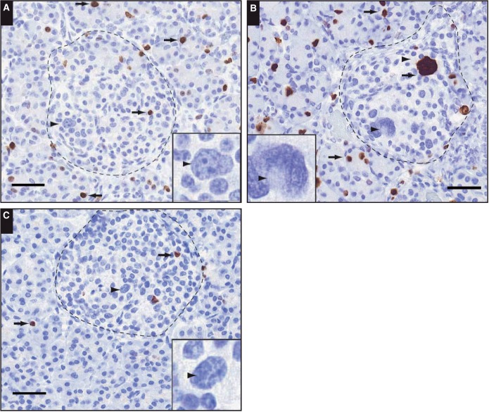
Objectives: To quantify islet cell nucleomegaly in controls and tissues obtained from patients with congenital hyperinsulinism in infancy (CHI) and to examine the association of nucleomegaly with proliferation.

Methods: High-content analysis of histologic sections and serial block-face scanning electron microscopy were used to quantify nucleomegaly.

Results: Enlarged islet cell nuclear areas were 4.3-fold larger than unaffected nuclei, and the mean nuclear volume increased to approximately threefold. Nucleomegaly was a normal feature of pediatric islets and detected in the normal regions of the pancreas from patients with focal CHI. The incidence of nucleomegaly was highest in diffuse CHI (CHI-D), with more than 45% of islets containing two or more affected cells. While in CHI-D nucleomegaly was negatively correlated with cell proliferation, in all other cases, there was a positive correlation.

Conclusions: Increased incidence of nucleomegaly is pathognomonic for CHI-D, but these cells are nonproliferative, suggesting a novel role in the pathobiology of this condition.

Keywords: Congenital hyperinsulinism; Hyperplasia; Hypoglycemia; Insulin; Islet; Nucleomegaly; Pancreas; Serial block-face scanning electron microscopy; β cell.


Figures

Image 1
Image 1
Typical images of nucleomegaly (arrowheads and insets) in islets (dotted lines) from tissue sections obtained from different patients with congenital hyperinsulinism in infancy (CHI): CHI-6 (A), CHI-4 (B), and a 10-month-old control, (C). Tissue sections were stained using the proliferation marker Ki-67, and typical Ki-67–positive cells are illustrated by the arrows. Note that not all Ki-67–positive nuclei are enlarged and that not all enlarged nuclei are positive for Ki-67. Scale bar is equivalent to 40 μm (insets, ×2.5).
Image 2
Image 2
Islet cells with nucleomegaly have an endocrine phenotype. A, Staining in a congenital hyperinsulinism in infancy (CHI) islet with nucleomegaly indicated by the arrowhead. Scale bar is equivalent to 40 μm. B, A series of images obtained by serial block-face scanning electron microscopy through adjacent parts of the tissue. Note the limited number of secretory granules (indicated by arrows) in the cell with nucleomegaly (N) compared with the surrounding cells and the control cell illustrated in the right-hand montage (C). Each image is separated by 1 μm. Scale bars are equivalent to 2 μm; expanded image 500 nm. The data were obtained from patients CHI-9 (A) and CHI-11 (B).
Image 2
Image 2
Islet cells with nucleomegaly have an endocrine phenotype. A, Staining in a congenital hyperinsulinism in infancy (CHI) islet with nucleomegaly indicated by the arrowhead. Scale bar is equivalent to 40 μm. B, A series of images obtained by serial block-face scanning electron microscopy through adjacent parts of the tissue. Note the limited number of secretory granules (indicated by arrows) in the cell with nucleomegaly (N) compared with the surrounding cells and the control cell illustrated in the right-hand montage (C). Each image is separated by 1 μm. Scale bars are equivalent to 2 μm; expanded image 500 nm. The data were obtained from patients CHI-9 (A) and CHI-11 (B).
Figure 1
Figure 1
Estimates of the surface area of nuclei in congenital hyperinsulinism in infancy and control cells. A, The ranges of nuclear areas from 393 cells; exocrine cells had a small range of nuclear areas compared with endocrine cells, and both were much smaller than the distribution of nuclear areas from islet cells exhibiting nucleomegaly. B, Enlarged nuclei in islet cells had an area of 101.1 ± 3.8 µm2 (mean ± SEM, n = 105) compared with 23.0 ± 0.4 μm2 (n = 173) and 18.7 ± 0.3 μm2 (n = 115) in control endocrine and exocrine cells, respectively. aP < .001.
Figure 2
Figure 2
Nuclear volume estimates in congenital hyperinsulinism in infancy (CHI) islet cells. A, Summary of the volume estimates of control nuclei (mean ± SEM; 162.32 ± 8 μm3, n = 30) and enlarged nuclei (452.83 ± 119.8 μm3, n = 4) in islet cells. The data were obtained from three patient samples with CHI. B, Transmission electron microscopy data and a digital reconstruction of control islet cell nuclei (C) and a cell exhibiting nucleomegaly (N), using serial block-face scanning electron microscopy. A total of 485 serial sections of tissue in 100-nm thicknesses were used to generate the data set. Scale bar is equivalent to 2 μm. Data obtained from patient CHI-11.
Figure 3
Figure 3
The incidence of nucleomegaly in islet cells. These panels summarize the range of enlarged nuclei observed in islet structures using tissue sections for each congenital hyperinsulinism in infancy (CHI) cohort and age-matched control tissue. The number of enlarged nuclei per islet surface area has been expressed relative to the number of cells within the islet. Only diffuse CHI (CHI-D) islets (B) had a high incidence of more than four enlarged nuclei per islet surface area. Indeed, some CHI-D islets were found to contain up to nine nucleomegalic cells in a single field of view. A, Control. B, CHI-D. C, Atypical CHI. D, Focal, control.
Image 3
Image 3
A-C, Serial sections of tissue to illustrate the presence of multiple enlarged nuclei, indicated by arrows. Note that most of the nuclei appear in successive 5-μm sections of tissue. Data were obtained from patient CHI-9. Scale bar is equivalent to 30 μm.
Figure 4
Figure 4
Multiple islet cell nucleomegaly in diffuse congenital hyperinsulinism in infancy (CHI-D) islets. An analysis of multiple enlarged nuclei in control (open shapes) and CHI-D islets (closed shapes). The comparative data set was obtained from a patient with congenital hyperinsulinism in infancy at two time points following two pancreatectomies—one at 13 months (squares) and the other at 30 months of age (circles). Fifty serial sections of tissue were used to assess total islet cell nucleomegaly from a minimum of 10 islets. Note how 100% of CHI-D islets had enlarged nuclei (n = 20). The minimum number of cells with nucleomegaly was six per islet, and one islet was found to contain 21 nucleomegalic cells. By comparison, fewer control islets contained nucleomegalic cells.

Similar articles

Cited by

References

    1. Dunne MJ, Cosgrove KE, Shepherd RM, et al. Hyperinsulinism in infancy: from basic science to clinical disease. Physiol Rev. 2004;84:239-275. - PubMed
    1. Glaser B. Lessons in human biology from a monogenic pancreatic β cell disease. J Clinical Invest. 2011;121:3821-3825. - PMC - PubMed
    1. Arya VB, Mohammed Z, Blankenstein O, et al. Hyperinsulinaemic hypoglycaemia. Horm Metab Res. 2014;46:157-170. - PubMed
    1. De Leon DD, Stanley CA. Mechanisms of disease: advances in diagnosis and treatment of hyperinsulinism in neonates. Nat Clin Pract Endocrinol Metab. 2007;3:57-68. - PubMed
    1. Yorifuji T. Congenital hyperinsulinism: current status and future perspectives. Ann Pediatr Endocrinol Metab. 2014;19:57-68. - PMC - PubMed