Volumetric capnography: lessons from the past and current clinical applications
- PMID: 27334879
- PMCID: PMC4918076
- DOI: 10.1186/s13054-016-1377-3
Volumetric capnography: lessons from the past and current clinical applications
Abstract
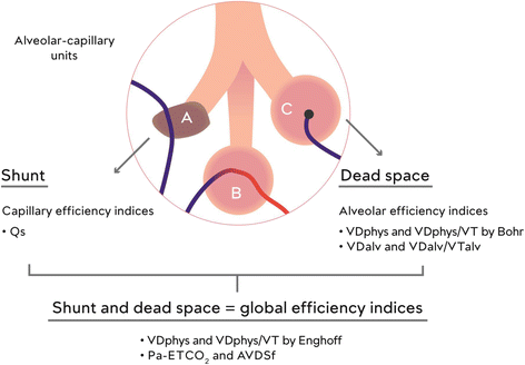
Dead space is an important component of ventilation-perfusion abnormalities. Measurement of dead space has diagnostic, prognostic and therapeutic applications. In the intensive care unit (ICU) dead space measurement can be used to guide therapy for patients with acute respiratory distress syndrome (ARDS); in the emergency department it can guide thrombolytic therapy for pulmonary embolism; in peri-operative patients it can indicate the success of recruitment maneuvers. A newly available technique called volumetric capnography (Vcap) allows measurement of physiological and alveolar dead space on a regular basis at the bedside. We discuss the components of dead space, explain important differences between the Bohr and Enghoff approaches, discuss the clinical significance of arterial to end-tidal CO2 gradient and finally summarize potential clinical indications for Vcap measurements in the emergency room, operating room and ICU.
Figures

Similar articles
-
Assessment of dead-space ventilation in patients with acute respiratory distress syndrome: a prospective observational study.Crit Care. 2016 May 5;20(1):121. doi: 10.1186/s13054-016-1311-8. Crit Care. 2016. PMID: 27145818 Free PMC article.
-
Assessment of Bohr and Enghoff Dead Space Equations in Mechanically Ventilated Children.Respir Care. 2017 Apr;62(4):468-474. doi: 10.4187/respcare.05108. Epub 2017 Feb 21. Respir Care. 2017. PMID: 28223465
-
Comparison of volumetric capnography and mixed expired gas methods to calculate physiological dead space in mechanically ventilated ICU patients.Intensive Care Med. 2012 Oct;38(10):1712-7. doi: 10.1007/s00134-012-2670-5. Epub 2012 Aug 15. Intensive Care Med. 2012. PMID: 22893221
-
Volume Capnography in the Intensive Care Unit: Physiological Principles, Measurements, and Calculations.Ann Am Thorac Soc. 2019 Mar;16(3):291-300. doi: 10.1513/AnnalsATS.201807-501CME. Ann Am Thorac Soc. 2019. PMID: 30657700 Review.
-
Volumetric capnography: the time has come.Curr Opin Crit Care. 2014 Jun;20(3):333-9. doi: 10.1097/MCC.0000000000000095. Curr Opin Crit Care. 2014. PMID: 24785676 Review.
Cited by
-
A mathematical model for the first derivative wave analysis of the volumetric capnogram from the perspective of erythrocyte motion profiles.Heliyon. 2019 Jun 12;5(6):e01824. doi: 10.1016/j.heliyon.2019.e01824. eCollection 2019 Jun. Heliyon. 2019. PMID: 31223667 Free PMC article.
-
Novel volumetric capnography indices measure ventilation inhomogeneity in cystic fibrosis.ERJ Open Res. 2022 Mar 14;8(1):00440-2021. doi: 10.1183/23120541.00440-2021. eCollection 2022 Jan. ERJ Open Res. 2022. PMID: 35295235 Free PMC article.
-
The effect of long-acting dual bronchodilator therapy on exercise tolerance, dynamic hyperinflation, and dead space during constant work rate exercise in COPD.J Appl Physiol (1985). 2021 Jun 1;130(6):2009-2018. doi: 10.1152/japplphysiol.00774.2020. Epub 2021 Apr 29. J Appl Physiol (1985). 2021. PMID: 33914661 Free PMC article. Clinical Trial.
-
Effects of 2 modes of positive pressure ventilation on respiratory mechanics and gas exchange in foals.J Vet Intern Med. 2023 May-Jun;37(3):1233-1242. doi: 10.1111/jvim.16651. Epub 2023 Apr 13. J Vet Intern Med. 2023. PMID: 37051768 Free PMC article.
-
Validation of a method for estimating pulmonary dead space in ventilated beagles to correct exhaled propofol concentration in mixed air.BMC Vet Res. 2025 Jan 7;21(1):9. doi: 10.1186/s12917-024-04458-1. BMC Vet Res. 2025. PMID: 39773486 Free PMC article.
References
-
- Fowler WS. Lung function studies; the respiratory dead space. Am J Physiol. 1948;154(3):405–16. - PubMed
Publication types
MeSH terms
LinkOut - more resources
Full Text Sources
Other Literature Sources
Medical
Miscellaneous

