. 2016 Aug 12;291(33):17437-49.
doi: 10.1074/jbc.M116.724013. Epub 2016 Jun 22.

Identification of a Conserved RNA-dependent RNA Polymerase (RdRp)-RNA Interface Required for Flaviviral Replication

Affiliations

Identification of a Conserved RNA-dependent RNA Polymerase (RdRp)-RNA Interface Required for Flaviviral Replication

Kenneth Hodge et al. J Biol Chem. .

Abstract

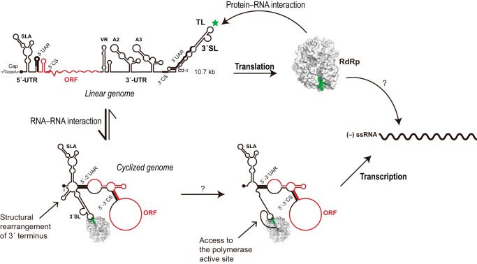
Dengue virus, an ∼10.7-kb positive-sense RNA virus, is the most common arthropod-communicated pathogen in the world. Despite dengue's clear epidemiological importance, mechanisms for its replication remain elusive. Here, we probed the entire dengue genome for interactions with viral RNA-dependent RNA polymerase (RdRp), and we identified the dominant interaction as a loop-forming ACAG motif in the 3' positive-stranded terminus, complicating the prevailing model of replication. A subset of interactions coincides with known flaviviral recombination sites inside the viral protein-coding region. Specific recognition of the RNA element occurs via an arginine patch in the C-terminal thumb domain of RdRp. We also show that the highly conserved nature of the consensus RNA motif may relate to its tolerance to various mutations in the interacting region of RdRp. Disruption of the interaction resulted in loss of viral replication ability in cells. This unique RdRp-RNA interface is found throughout flaviviruses, implying possibilities for broad disease interventions.

Keywords: RNA-dependent RNA polymerase (RdRp); dengue virus (DENV); untranslated region (UTR); viral non-structural protein (NS) 5; viral polymerase; viral protein; viral replication; virology; yeast three-hybrid (Y3H).


Figures

FIGURE 1.
FIGURE 1.
Interaction landscape of DENV RdRp with the entire RNA genome. A, schematic diagram of Y3H in this study. B, Western blotting with polyclonal anti-NS5 antibodies (Gentex 103350) confirmed the expression of DENV-2 NS5 (120.9 kDa) and its RdRp (90.1 kDa) in Y3H with two representative RNAs, SLA and the full-length 5′-UTR. The protein IRP in pACT2 was used as a negative control. C, DENV genome is depicted as light gray rectangles and is numbered. The coding regions for viral proteins are labeled. Positive strand RNA fragments of the genome that bind the full-length NS5 and RdRp are shown above the genome as black and gray rectangles, respectively, whereas negative fragments are below. Levels of yeast growth in the presence of 3-AT are indicated by three symbols. Representative Mfold structures of RNA fragments are drawn with the CACAG motif in red. D, conservation of the ACAG loop motif is compared between the top loop regions of the 5′-SLA and the 3′-SL. Sequences are ordered according to a phylogenetic tree based on the terminal 120 nucleotides of Flaviviridae. The left vertical bar indicates the type of Flaviviridae as follows: black indicates mosquito-borne viruses; white indicates tick-borne viruses; light gray indicates viruses of no known vector; and dark gray indicates a single hepacivirus example. Consensus nucleotides are indicated once at the bottom of the figure; elsewhere, they are indicated by hyphens, with variations from the consensus indicated by the appropriate letter.
FIGURE 2.
FIGURE 2.
DENV RdRp recognizes the top loop of 3′-SL. Linear 3′-SL and a series of its deletion mutants are indicated in the secondary structure with rectangles (upper left panel), whereas the model of the cyclized Y3H RNA construct containing DENV 5′- and 3′-terminal regions (cyUTR) used in this study is in the upper right panel. The long viral open reading frame was replaced by a GNRA loop that linked 5′- and 3′-UTR together in the cyUTR construct. SL mutant ΔsHP completely removed the boxed hairpin; ΔS1 removed the lower SL stem; ΔS2 removed the upper SL stem; ΔTL removed the indicated top loop nucleotides; ΔSiL removed the side loop; and ΔTail replaced the final 5′ nucleotides with CAAAA. In cyUTR, SLAmut replaced the wild-type GAU with CG, and U>C refers to the substitution of a U with a single C. Structural RNA elements are labeled. β-Galactosidase expression assays showing the interaction strengths with the full-length NS5 (black-filled bar) or RdRp (gray) are measured in Miller units. The lower left panel shows RdRp interactions with a variety of linear DENV RNAs with native sequences (left half) and mutations (right half). Results with a cyclized form of UTR are separated into the lower right panel. Error bars indicate S.E.
FIGURE 3.
FIGURE 3.
In vitro EMSA. Mfold secondary structures of RNAs are depicted in the left column. Arrows highlight protein-RNA complexes that were evident via both EtBr (middle column) and Coomassie (right column) staining. The constant amount of RNA in the reaction was increased from 37.5 to 50 pmol in the case of protein staining. The linSL and cirSL experiments were run on a single gel with one “RdRp alone” lane.
FIGURE 4.
FIGURE 4.
In vitro Alpha binding assay. A, saturation binding experiment of RdRp with truncated 3′-SL. The Alpha-binding signal is shown in fluorescence units (FU). B, competition assay with unlabeled RNA. RdRp and Bi-miniSL concentrations were kept constant at 200 and 10 nm, respectively. Mean and standard deviation values are derived from three independent experiments. We note that SLA did not show a binding signal in both saturation binding and competition assay.
FIGURE 5.
FIGURE 5.
Exploration of 3′-SL-binding sites in RdRp using Y3H random mutagenesis. A, random RdRp mutants retaining SL-binding capacity were sequenced and analyzed as under “Experimental Procedures.” Results show two specific regions of interest in the thumb domain (dark gray), which were highly susceptible to mutations (stars). White areas indicate the finger domain and light gray shows the palm domain. B, mutations allowing RdRp binding in Y3H are mapped onto the crystal structure (Protein Data Bank code 2J7W) in the left panel (violet), together with naturally occurring variations found in 2,446 DENV sequences from the NCBI database (cyan). Residues overlapping between the two data sets are shown in yellow. In the middle panel, the protein solvent-accessible surfaces were generated with the program APBS handled in PyMOL and are colored according to their electrostatic potential from red to blue. Our results prompted us to manually create an RNA-docking model using YFV SL structures (Protein Data Bank code 2KPC) (right panel). RdRP and SL-TL were manually docked on PyMOL to minimize steric clashes and to reproduce mutational studies. C, Miller assays for SL interactions with specific RdRp mutants are shown (left panel), together with their expression in yeast (right panel). We have no clear explanation for the double banding pattern that was occasionally observed. D, DENV replicon assay with RdRp mutants in BHK-21 cells. Viral replication was investigated via YFP signals 4 days after transfection to monitor RNA synthesis. YFP signals are shown for each mutant. ΔTL refers to mutations that altered the 3′-SL top loop to the extent that RdRp binding was abrogated in Y3H (Fig. 2). GAA refers to an inactive RdRp by mutation of its catalytic triad, GDD.
FIGURE 6.
FIGURE 6.
Exploring the conservation of the RdRp-RNA interface within and beyond DENV. A, heat map matrix of interaction strengths among RdRp and 3′-SL variants. Relative interaction strengths of 3′-SL variants with the DENV RdRp are shown in a box as a percentage of the wild-type (WT) RdRp-CACAG pair. Colors, indicating a 3′-SL-TL variant's sensitivity to protein alterations, were arrived at as follows: % changes in interaction strengths between mutant RdRp and 3′-SL variant combinations versus those of WT RdRp and 3′-SL variants are calculated. These percentage changes are then compared (via simple subtraction) to the relevant RdRp-CACAG interaction strengths. Numerous RNA variants, including some residing on the 5′-SLA, are not included on the table, as their interactions with RdRp were particularly weak. B, analysis of 3′-SL-TL's tolerance for mutations in Arg-773. A library of Arg-773 RdRp mutants (R773X) was examined via Y3H with five 3′-SL-TL variants under one permissive and two selective conditions. C, DALI structural alignments of the thumb domains from five available Flaviviridae RdRp structures. Residues with perfect conservation are colored in red, and ones that align in four sequences are green. The null symbols (ø) indicate mutations that had no effect in 3′-SL-binding in our Y3H random mutagenesis. Filled triangles point to residues that were chosen for site-directed mutagenesis, and red triangles indicate crucial residues in 3′-SL-TL-binding. HCV residues making contacts with thumb 2 inhibitors in previous studies are highlighted by blue lines. The gray rectangle shows the region that was deleted in an HCV structural study (45). D, superposition of five Flaviviridae RdRp structures reveals similarity in the SL-binding site of DENV RdRp comprising an Arg-770–Tyr-838–Arg-773 stacking platform. In all cases, two non-contiguous helices combine to form a positively charged pocket (blue) centering on a perfectly conserved Arg-773 in DENV.
FIGURE 7.
FIGURE 7.
Schematic model of DENV replication based on our results coupling with earlier studies. We propose that the structural rearrangements in both polymerase and the 3′ end region of genomic RNA upon binding of RdRp to the 3′-SL-TL and genomic cyclization play key roles in regulation of viral replication cycle. Accurate mechanisms for recruitment of the 3′ end into the active site of RdRp as well as recognition of the negative stranded genome by RdRp still remain elusive.

Similar articles

Cited by

References

    1. Dreher T. W., and Hall T. C. (1988) Mutational analysis of the sequence and structural requirements in brome mosaic virus RNA for minus strand promoter activity. J. Mol. Biol. 201, 31–40 - PubMed
    1. Song C., and Simon A. E. (1995) Requirement of a 3′-terminal stem-loop in in vitro transcription by an RNA-dependent RNA polymerase. J. Mol. Biol. 254, 6–14 - PubMed
    1. Oh J. W., Sheu G. T., and Lai M. M. (2000) Template requirement and initiation site selection by hepatitis C virus polymerase on a minimal viral RNA template. J. Biol. Chem. 275, 17710–17717 - PubMed
    1. Cui T., and Porter A. G. (1995) Localization of binding site for encephalomyocarditis virus RNA polymerase in the 3′-noncoding region of the viral RNA. Nucleic Acids Res. 23, 377–382 - PMC - PubMed
    1. Filomatori C. V., Lodeiro M. F., Alvarez D. E., Samsa M. M., Pietrasanta L., and Gamarnik A. V. (2006) A 5′ RNA element promotes dengue virus RNA synthesis on a circular genome. Genes Dev. 20, 2238–2249 - PMC - PubMed

MeSH terms