Review
. 2016 Jul;96(3):1071-91.
doi: 10.1152/physrev.00035.2015.

The Function of V-ATPases in Cancer

Affiliations
Review

The Function of V-ATPases in Cancer

Laura Stransky et al. Physiol Rev. 2016 Jul.

Abstract

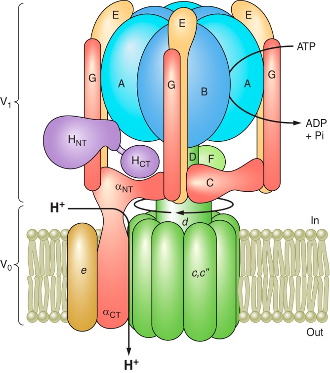
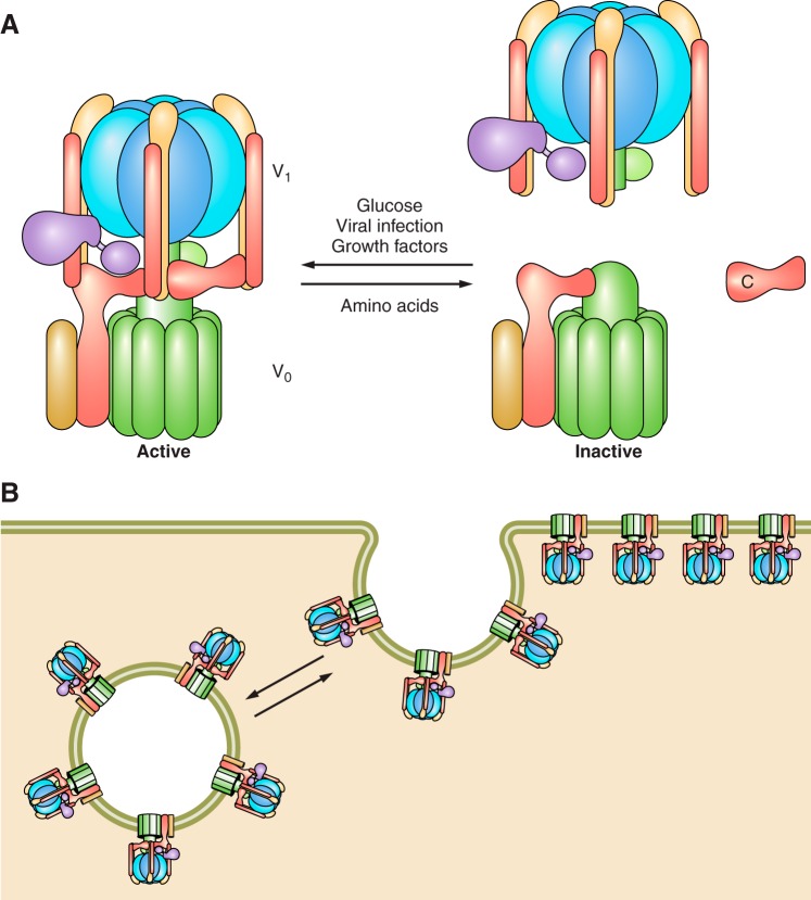
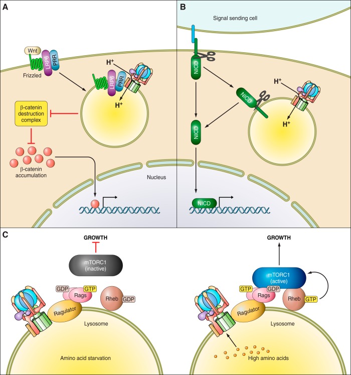
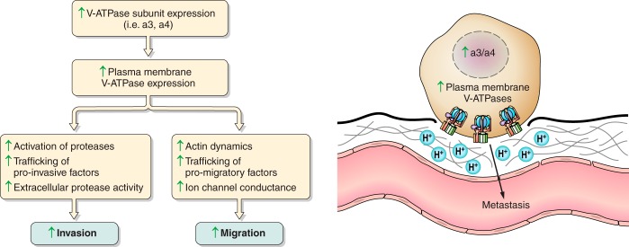
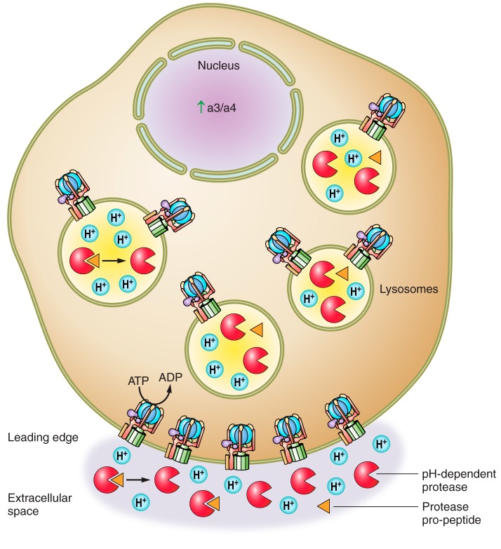
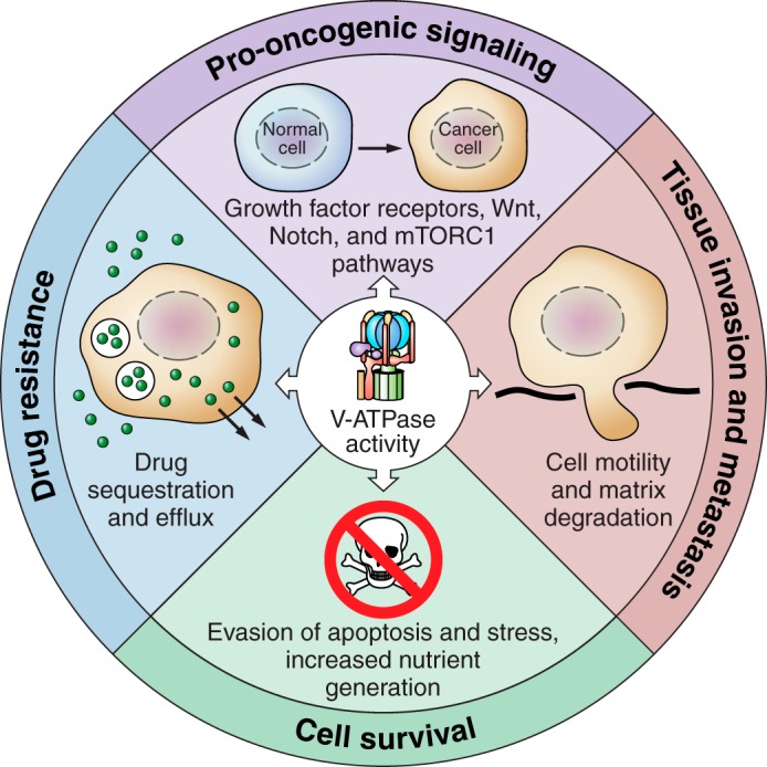
The vacuolar ATPases (V-ATPases) are a family of proton pumps that couple ATP hydrolysis to proton transport into intracellular compartments and across the plasma membrane. They function in a wide array of normal cellular processes, including membrane traffic, protein processing and degradation, and the coupled transport of small molecules, as well as such physiological processes as urinary acidification and bone resorption. The V-ATPases have also been implicated in a number of disease processes, including viral infection, renal disease, and bone resorption defects. This review is focused on the growing evidence for the important role of V-ATPases in cancer. This includes functions in cellular signaling (particularly Wnt, Notch, and mTOR signaling), cancer cell survival in the highly acidic environment of tumors, aiding the development of drug resistance, as well as crucial roles in tumor cell invasion, migration, and metastasis. Of greatest excitement is evidence that at least some tumors express isoforms of V-ATPase subunits whose disruption is not lethal, leading to the possibility of developing anti-cancer therapeutics that selectively target V-ATPases that function in cancer cells.


Figures

FIGURE 1.
FIGURE 1.
Structure and mechanism of the V-ATPase. The V-ATPase is composed of a peripheral V1 domain that hydrolyzes ATP and an integral V0 domain that translocates protons. ATP hydrolysis occurs at nucleotide binding sites located at the interface of the A and B subunits and drives rotation of a central rotary complex composed of subunits D and F of V1 and subunit d and the ring of proteolipid subunits (c and c”) of V0 relative to the remainder of the complex. Rotation of the proteolipid ring relative to subunit a drives unidirectional proton transport from the cytoplasm to the lumen (see text for details). The A3B3 catalytic head is held fixed relative to subunit a by peripheral stalks composed of three EG heterodimers that connect to subunits C and H and the NH2-terminal cytoplasmic domain of subunit a. Model is adapted from Couoh-Cardel et al. (26). See Reference 232 for a recent model based on cryo-EM of the yeast V-ATPase.
FIGURE 2.
FIGURE 2.
Regulation of V-ATPase activity. A: V-ATPase activity is regulated in vivo by reversible dissociation of the complex into its component V1 and V0 domains, which results in inactivation of the complex. In mammalian cells, regulated assembly occurs in response to a number of cues, including changes in glucose concentration, starvation of amino acids, exposure to growth factors, upon maturation of dendritic cells, and during infection of cells by influenza virus. PI3K is involved in controlling assembly in response to changes in glucose concentration, during dendritic cell maturation and during viral infection, but not in response to changes in amino acid levels. B: V-ATPase activity in epithelial cells in the kidney and epididymis is controlled by regulated trafficking of complexes to the apical membrane of these cells. In both systems, trafficking is controlled by PKA downstream of a bicarbonate-sensitive adenylate cyclase.
FIGURE 3.
FIGURE 3.
The V-ATPase is required for cellular signaling. A: the role of the V-ATPase in Wnt signaling. Wnt ligand binds to the plasma membrane receptor Frizzled in complex with a coreceptor, here LRP6. LRP6 and Frizzled are bound by the prorenin receptor (PRR), which acts as an adapter between the receptor complex and the V-ATPase. Upon ligand binding, the complex must be internalized into a signaling endosome for full activation via LRP6 phosphorylation, which requires V-ATPase activity. This activation inhibits activity of the β-catenin destruction complex, allowing β-catenin to accumulate, translocate to the nucleus, and alter gene transcription. B: the role of the V-ATPase in Notch signaling. The Notch receptor is activated by binding of Notch ligand expressed on the surface of an adjacent cell. Ligand binding stimulates a series of proteolytic cleavages which release the Notch intracellular domain (NICD) from the membrane, allowing it to be imported into the nucleus and drive transcription of Notch target genes. The cleavage of the Notch receptor is enhanced by V-ATPase-dependent acidification of endosomes after receptor internalization. C: the role of the V-ATPase in mTORC1 signaling. The V-ATPase is part of the amino acid sensing machinery associated with the surface of the lysosome. In the absence of amino acids (left panel), the V-ATPase is tightly complexed with the Ragulator, and the RagGTPase heterodimer is inactive. As a result, mTORC1 is inactive in the cytosol, thus repressing anabolic processes and cellular growth. When amino acids are sufficient in the cell (right panel), the V-ATPase-Ragulator complex undergoes a conformational change, stimulating Ragulator to act as a GEF, activating the RagGTPases. The active Rags recruit mTORC1 from the cytosol to the lysosomal surface where it is activated by Rheb. Active mTORC1 then promotes cellular growth.
FIGURE 4.
FIGURE 4.
The promotion of metastasis via plasma membrane V-ATPase activity. Upregulation of V-ATPase subunits such as a3 or a4 localize V-ATPases to the plasma membrane, where they participate in a number of processes that enhance cancer cell invasion and migration. Plasma membrane V-ATPases are believed to activate pH-dependent proteases, promote trafficking of pro-invasive factors, and enhance extracellular protease activity. They are also thought to interact with and alter actin dynamics, enhance trafficking of pro-migratory factors, and alter ion channel conductance to promote migration. Enhanced invasion and migration of cancer cells eventually leads to the escape of tumor cells from the primary tumor site and the development of metastases.
FIGURE 5.
FIGURE 5.
Mechanisms by which the V-ATPase enhances tumor cell invasiveness and survival. The primary mechanism by which the V-ATPase is thought to promote tumor cell invasion is through the promotion of extracellular pH-dependent protease activity. Intracellularly, V-ATPase-mediated acidification of intracellular compartments may allow for activation of proteases that are then trafficked to the cell surface. V-ATPases localized to the plasma membrane of cancer cells upon upregulation of the subunit a isoforms a3 or a4 may create an acidic microenvironment that allows for activation of pH-dependent proteases and/or enhances pH-dependent protease activity outside of the cell. Enhanced plasma membrane V-ATPase activity also increases acid secretion out of the cytosol. This is important for tumor cell survival as tumor cells generate large amounts of metabolic acid due to their reliance on glycolytic metabolism and are able to survive in highly acidic microenvironments.
FIGURE 6.
FIGURE 6.
The role of V-ATPases in cancer. V-ATPase activity supports pro-oncogenic signaling pathways, including growth factor receptors, Wnt, Notch, and mTORC1 (top panel). V-ATPase activity allows tumor cells to evade apoptosis and various cellular stressors (right panel). V-ATPase activity promotes the ability of tumor cells to invade surrounding tissue and metastasize to secondary sites within the body by promoting cell motility and extracellular matrix degradation (bottom panel). V-ATPase activity contributes to drug resistance by promoting drug trapping in acidic cellular spaces and promoting drug efflux out of the cell (left panel).

References

    1. Adams DS, Masi A, Levin M. H+ pump-dependent changes in membrane voltage are an early mechanism necessary and sufficient to induce Xenopus tail regeneration. Development 134: 1323–1335, 2007. - PubMed
    1. Adams DS, Robinson KR, Fukumoto T, Yuan S, Albertson RC, Yelick P, Kuo L, McSweeney M, Levin M. Early, H+-V-ATPase-dependent proton flux is necessary for consistent left-right patterning of non-mammalian vertebrates. Development 133: 1657–1671, 2006. - PMC - PubMed
    1. Alwan HAJ, van Zoelen EJJ, van Leeuwen JEM. Ligand-induced lysosomal epidermal growth factor receptor (EGFR) degradation is preceded by proteasome-dependent EGFR de-ubiquitination. J Biol Chem 278: 35781–35790, 2003. - PubMed
    1. Alzamora R, Thali RF, Gong F, Smolak C, Li H, Baty CJ, Bertrand CA, Auchli Y, Brunisholz RA, Neumann D, Hallows KR, Pastor-Soler NM. PKA regulates vacuolar H+-ATPase localization and activity via direct phosphorylation of the a subunit in kidney cells. J Biol Chem 285: 24676–24685, 2010. - PMC - PubMed
    1. Amsterdam A, Nissen RM, Sun Z, Swindell EC, Farrington S, Hopkins N. Identification of 315 genes essential for early zebrafish development. Proc Natl Acad Sci USA 101: 12792–12797, 2004. - PMC - PubMed

Publication types

MeSH terms

Substances

LinkOut - more resources