. 2016 Jul;28(7):1738-51.
doi: 10.1105/tpc.15.00870. Epub 2016 Jun 22.

Multiple Calmodulin-Binding Sites Positively and Negatively Regulate Arabidopsis CYCLIC NUCLEOTIDE-GATED CHANNEL12

Affiliations

Multiple Calmodulin-Binding Sites Positively and Negatively Regulate Arabidopsis CYCLIC NUCLEOTIDE-GATED CHANNEL12

Thomas A DeFalco et al. Plant Cell. 2016 Jul.

Abstract

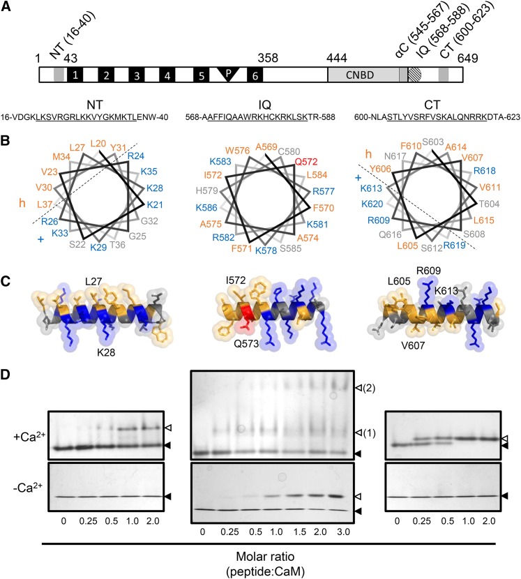
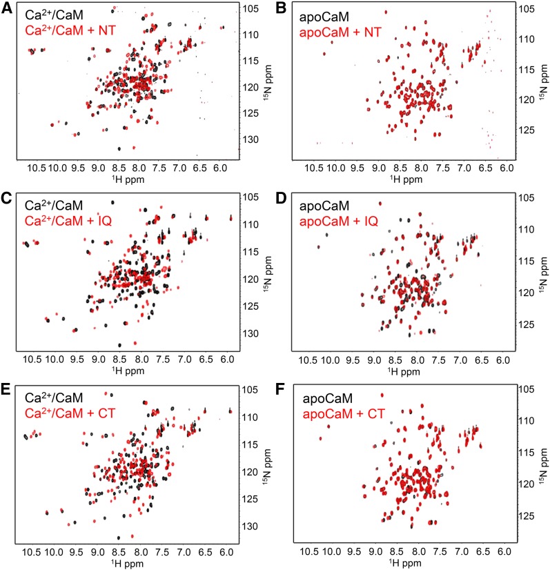
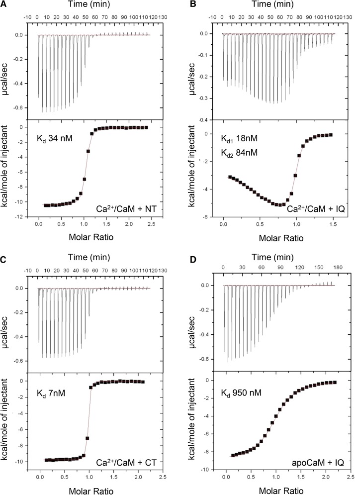
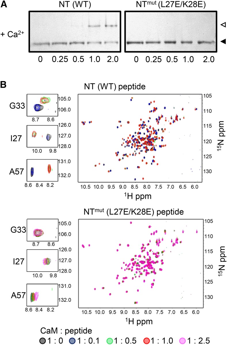
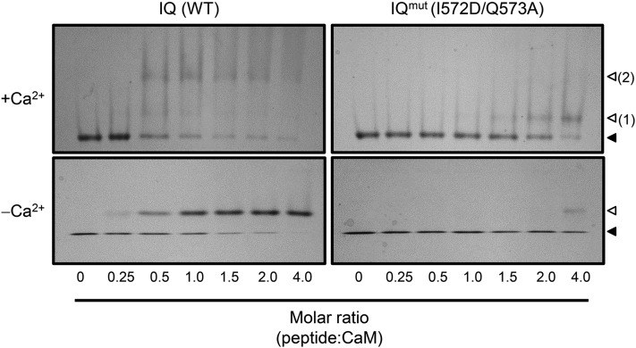
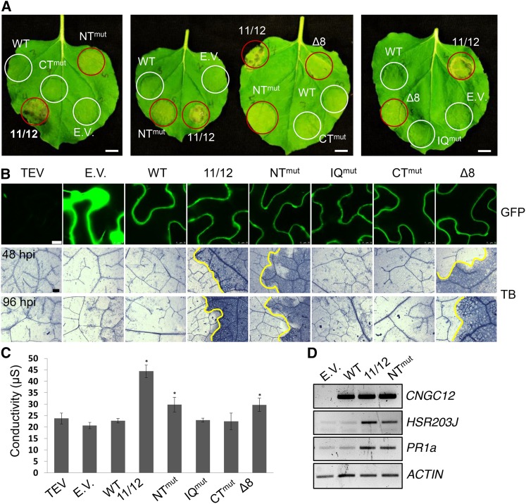
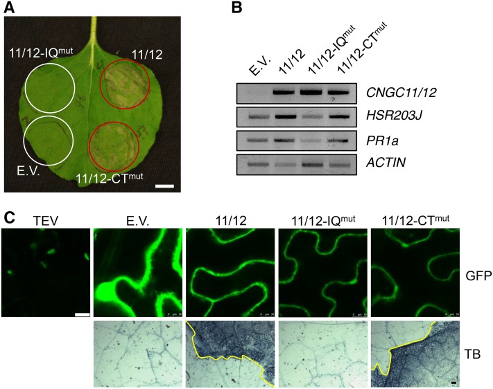
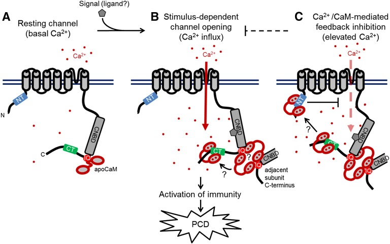
Ca(2+) signaling is critical to plant immunity; however, the channels involved are poorly characterized. Cyclic nucleotide-gated channels (CNGCs) are nonspecific, Ca(2+)-permeable cation channels. Plant CNGCs are hypothesized to be negatively regulated by the Ca(2+) sensor calmodulin (CaM), and previous work has focused on a C-terminal CaM-binding domain (CaMBD) overlapping with the cyclic nucleotide binding domain of plant CNGCs. However, we show that the Arabidopsis thaliana isoform CNGC12 possesses multiple CaMBDs at cytosolic N and C termini, which is reminiscent of animal CNGCs and unlike any plant channel studied to date. Biophysical characterizations of these sites suggest that apoCaM interacts with a conserved isoleucine-glutamine (IQ) motif in the C terminus of the channel, while Ca(2+)/CaM binds additional N- and C-terminal motifs with different affinities. Expression of CNGC12 with a nonfunctional N-terminal CaMBD constitutively induced programmed cell death, providing in planta evidence of allosteric CNGC regulation by CaM. Furthermore, we determined that CaM binding to the IQ motif was required for channel function, indicating that CaM can both positively and negatively regulate CNGC12. These data indicate a complex mode of plant CNGC regulation by CaM, in contrast to the previously proposed competitive ligand model, and suggest exciting parallels between plant and animal channels.


Figures

Figure 1.
Figure 1.
Structural Modeling and ND-PAGE Mobility Shift Assay of CNGC12 CaMBD Peptides. (A) Location of the CaMBDs of CNGC12 (see delineation of NT and CT sites in Supplemental Figure 1). Numbers indicate amino acid position. 1 to 6, Transmembrane helices; P, pore region. (B) Helical wheel projections of motifs for the NT, IQ, and CT peptides (underlined sequences). Dashed lines separate proposed hydrophobic (h) and basic (+) faces of the NT and CT wheels. (C) 3D model of full-length CaMBD peptides. Specific residues used for mutant analysis of each motif in this work are indicated in each model. In both (B) and (C), hydrophobic and basic residues are colored orange and blue, respectively, while the Q573 residue of the IQ motif is also colored (red) due to its suspected involvement in CaM binding. (D) ND-PAGE mobility shift assays in the presence of 0.1 mM CaCl2 (top panels) or 2 mM EGTA (bottom panels). Closed triangles indicate the migration size of CaM alone, while open triangles indicate the migration size of CaM-peptide complex. Two distinct IQ-CaM complexes consistently migrated separately in Ca2+/CaM assays with IQ peptide (top panel, numbered 2); such a pattern was never observed with NT or CT peptides or in any apoCaM assays.
Figure 2.
Figure 2.
HSQC-NMR Spectroscopic Analysis of CaM-Peptide Interactions. Overlaid spectra of uniformly 15N-labeled CaM in the absence (black) or presence (red) of equimolar peptide. In all assays, spectra of 0.2 mM 15N-CaM were collected in either Ca2+ buffer (10 mM HEPES, 100 mM NaCl, and 5 mM CaCl2, pH 7.5) or apo buffer (10 mM HEPES, 100 mM NaCl, and 1 mM EGTA, pH 7.5), as indicated. Spectra of Ca2+/CaM (A) or apoCaM +/− NT peptide (B). Spectra of Ca2+/CaM (C) or apoCaM +/− IQ peptide (D). Spectra of Ca2+/CaM (E) or apoCaM +/− CT peptide (F).
Figure 3.
Figure 3.
ITC Analysis of Peptide-CaM Interactions. For all experiments, calorimetric titrations (top panels) and the least-squares fitted model of binding are shown (bottom panels). Calculated Kd values are shown for each modeled binding site. (A) Titration of 150 µM NT peptide into 16 µM CaM in the presence of 5 mM CaCl2. (B) Titration of 205 µM IQ peptide into 20 µM CaM in the presence of 5 mM CaCl2. (C) Titration of 150 µM CT peptide into 16 µM CaM in the presence of 5 mM CaCl2. (D) Titration of 138 µM IQ peptide into 20 µM CaM in the presence of 1 mM EGTA.
Figure 4.
Figure 4.
The L27E/K28E Mutation Disrupts CaM Binding to the CNGC12 NT Motif. (A) ND-PAGE of Ca2+/CaM with NT peptide or a peptide bearing the double mutation L27E/K28E (NTmut) at molar ratios indicated. Closed triangles indicate the migration size of CaM alone, while open triangles indicate the migration size of CaM-peptide complex. (B) Overlaid 1H-15N HSQC-NMR spectra of 0.2 mM uniformly 15N-labeled CaM with increasing molar ratios (as indicated under the figure) of NT (top) or NTmut (bottom) peptide. Spectra of 0.2 mM uniformly 15N-labeled CaM in the presence of increasing molar ratios of NTmut peptide. NMR samples were prepared in 10 mM Tris-Cl, 150 mM NaCl, and 10 mM CaCl2 pH 7.0. The peaks corresponding to three individual CaM residues (Gly-33, Ile-27, and Ala-57) are shown in enlarged panels.
Figure 5.
Figure 5.
The I572D/Q573A Mutation Disrupts CaM Binding to the CNGC12 IQ Motif. ND-PAGE was performed with IQ (wild-type sequence) or IQmut peptide (bearing the I672D/Q573A mutation). Closed and open arrows indicate the migration of free or peptide-bound CaM, respectively, while the two distinct bands formed by the IQ-Ca2+/CaM complex are numbered 1 and 2.
Figure 6.
Figure 6.
Transient Expression of NT Mutants Induces PCD in N. benthamiana. (A) Appearance of N. benthamiana leaves at 4 dpi (bar = 1 cm). Areas were infiltrated with Agrobacterium carrying different constructs. Areas showing cell death are circled in red. (B) GFP confocal microscopy of leaf areas 30 hpi (bar = 10 µm) and Trypan blue staining (TB) for cell death of N. benthamiana leaves expressing wild-type or mutant CNGC12 constructs at 48 and 96 hpi, as indicated (bar = 0.1 mm). Infiltrated and uninfiltrated leaf areas are separated by a yellow line in samples showing cell death. (C) Ion leakage of infiltrated leaf areas 4 dpi. Values shown are averages of three replicates (error bars = sd; *P < 0.05 compared with empty vector, Student’s t test). E.V., empty vector (GFP); WT, At-CNGC12; 11/12, CNGC11/12; NTmut, CNGC12L27E/K28E; IQmut, CNGC12I572D/Q573A; CTmut, CNGC12V607E; Δ8, CNGC12Δ32-39. (D) RT-PCR of cDNA from N. benthamiana leaves 24 hpi expressing wild-type or mutant CNGC12 constructs. Nb-ACTIN was used as a loading control (26 cycles).
Figure 7.
Figure 7.
The IQ Motif Is Required for CNGC11/12-Induced PCD. (A) N. benthamiana leaf 4 dpi (bar = 1 cm). (B) RT-PCR of cDNA from N. benthamiana leaves 24 hpi expressing CNGC11/12 or site-directed mutant constructs. Nb-ACTIN was used as a loading control (26 cycles). (C) GFP confocal microscopy of N. benthamiana leaves 30 hpi (bar = 10 µm) and Trypan blue (TB) staining for cell death in N. benthamiana leaves 3 dpi (bar = 0.1 mm). E.V., empty vector (GFP); 11/12, CNGC11/12; 11/12-IQmut, CNGC11/12I564D/Q565A; 11/12-CTmut, CNGC11/12V599E.
Figure 8.
Figure 8.
Proposed Model of CNGC12 Regulation by CaM. The model draws from the biophysical and physiological results of this study, previous data cited in the Discussion, and speculative components that currently remain unexplored, but which are compatible with our findings to date. (A) At resting conditions and basal Ca2+ levels, the channel is calmodulated via interaction between CaM and the IQ motif. (B) Upon perception of a signal (the molecular nature of which remains to be empirically determined), the channel opens and cytosolic Ca2+ levels increase. Given the necessity of CaM binding to the IQ motif for channel function as well as the higher-order interactions that occur between the IQ motif and Ca2+/CaM, channel activation may require CaM-dependent interactions between adjacent channel subunits as depicted. Furthermore, current data do not resolve whether individual CaM molecules move between different CaMBDs in a Ca2+-dependent manner or multiple CaMs can bind sites independently. (C) We propose that at higher Ca2+ concentration, Ca2+/CaM provides inhibition of CNGC12 function via the NT CaMBD; whether this inhibition involves the bridging of channel termini remains unclear.

References

    1. Abdel-Hamid H., Chin K., Moeder W., Shahinas D., Gupta D., Yoshioka K. (2013). A suppressor screen of the chimeric AtCNGC11/12 reveals residues important for intersubunit interactions of cyclic nucleotide-gated ion channels. Plant Physiol. 162: 1681–1693. - PMC - PubMed
    1. Arazi T., Kaplan B., Fromm H. (2000). A high-affinity calmodulin-binding site in a tobacco plasma-membrane channel protein coincides with a characteristic element of cyclic nucleotide-binding domains. Plant Mol. Biol. 42: 591–601. - PubMed
    1. Arazi T., Sunkar R., Kaplan B., Fromm H. (1999). A tobacco plasma membrane calmodulin-binding transporter confers Ni2+ tolerance and Pb2+ hypersensitivity in transgenic plants. Plant J. 20: 171–182. - PubMed
    1. Baxter J., Moeder W., Urquhart W., Shahinas D., Chin K., Christendat D., Kang H.G., Angelova M., Kato N., Yoshioka K. (2008). Identification of a functionally essential amino acid for Arabidopsis cyclic nucleotide gated ion channels using the chimeric AtCNGC11/12 gene. Plant J. 56: 457–469. - PubMed
    1. Ben Johny M., Yang P.S., Bazzazi H., Yue D.T. (2013). Dynamic switching of calmodulin interactions underlies Ca2+ regulation of CaV1.3 channels. Nat. Commun. 4: 1717. - PMC - PubMed

Publication types

MeSH terms

Grants and funding

LinkOut - more resources