. 2016 Mar 30:5:416.
doi: 10.12688/f1000research.8154.2. eCollection 2016.

Monoclonal antibodies against muscle actin isoforms: epitope identification and analysis of isoform expression by immunoblot and immunostaining in normal and regenerating skeletal muscle

Affiliations

Monoclonal antibodies against muscle actin isoforms: epitope identification and analysis of isoform expression by immunoblot and immunostaining in normal and regenerating skeletal muscle

Christine Chaponnier et al. F1000Res. .

Abstract

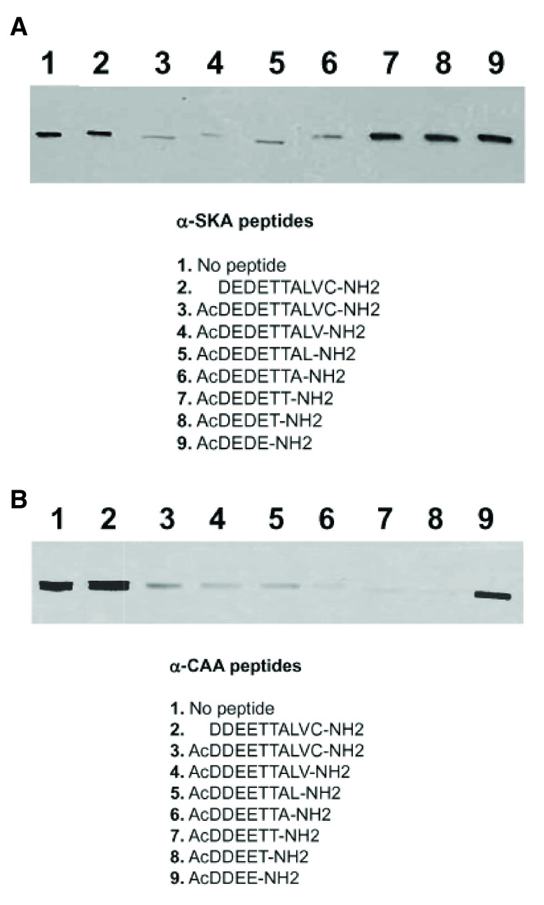
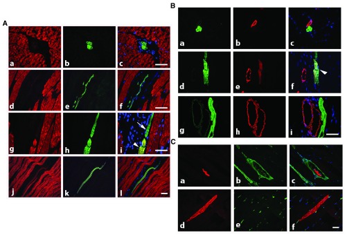
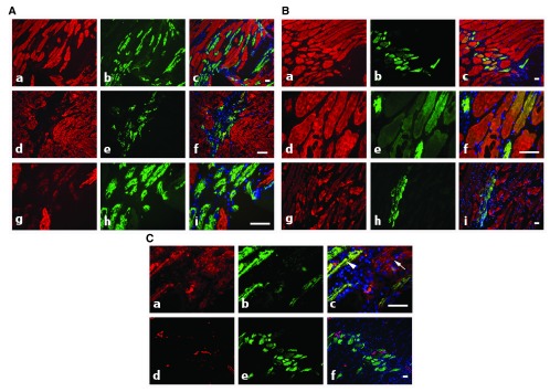
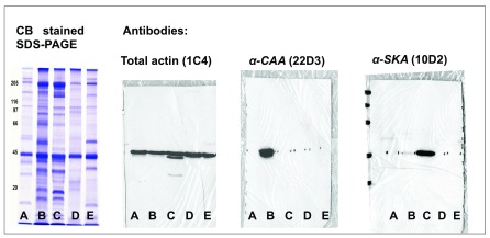
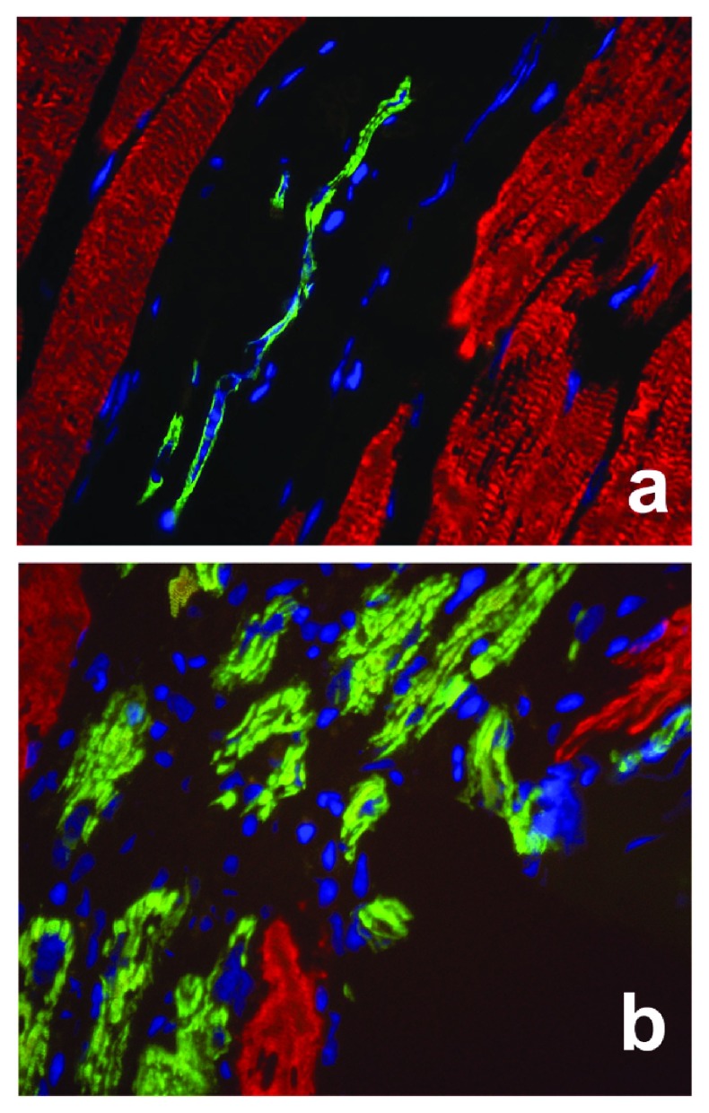
Higher vertebrates (mammals and birds) express six different highly conserved actin isoforms that can be classified in three subgroups: 1) sarcomeric actins, α-skeletal (α-SKA) and α-cardiac (α-CAA), 2) smooth muscle actins (SMAs), α-SMA and γ-SMA, and 3) cytoplasmic actins (CYAs), β-CYA and γ-CYA. The variations among isoactins, in each subgroup, are due to 3-4 amino acid differences located in their acetylated N-decapeptide sequence. The first monoclonal antibody (mAb) against an actin isoform (α-SMA) was produced and characterized in our laboratory in 1986 (Skalli et al., 1986) . We have further obtained mAbs against the 5 other isoforms. In this report, we focus on the mAbs anti-α-SKA and anti-α-CAA obtained after immunization of mice with the respective acetylated N-terminal decapeptides using the Repetitive Immunizations at Multiple Sites Strategy (RIMMS). In addition to the identification of their epitope by immunoblotting, we describe the expression of the 2 sarcomeric actins in mature skeletal muscle and during muscle repair after micro-lesions. In particular, we analyze the expression of α-CAA, α-SKA and α-SMA by co-immunostaining in a time course frame during the muscle repair process. Our results indicate that a restricted myocyte population expresses α-CAA and suggest a high capacity of self-regeneration in muscle cells. These antibodies may represent a helpful tool for the follow-up of muscle regeneration and pathological changes.

Keywords: Actin isoforms; epitope; monoclonal antibodies; muscle repair.


Conflict of interest statement

Competing interests: The authors sell the anti-actin isoform antibodies to companies through Unitec, University of Geneva, unitec@unige.ch

Figures

Figure 1.
Figure 1.
Identification, by immunoblots, of the epitope recognized by anti-α-SKA ( A) and anti-α-CAA ( B). Purified α-SKA ( A) or α-CAA ( B) was run on 10% SDS-PAGE and transferred on nitrocellulose membrane. Membrane strips were incubated with the mAb anti-α-SKA ( A) or anti-α-CAA ( B) alone (control, lane 1) or mixed with the listed peptides (2–9). The epitope recognized by anti-α-SKA ( A) includes the acetyl group and the first 7 amino acids of the α-SKA sequence. The epitope recognized by anti-α-CAA ( B) includes the acetyl group and the first 5 amino acids of the α-CAA sequence.
Figure 2.
Figure 2.. Rat adult skeletal muscle co-stained with isoform specific antibodies.
A) Co-staining with anti-α-SKA (red) and anti-α-CAA (green) allows the detection of isolated thin α-CAA positive fibers in interstitial connective tissue (transversal sections, a–c: longitudinal section, d–f) and of regenerating muscle fibers expressing both isoforms (longitudinal section, g–l, arrowhead), or only α-CAA (arrow). Merged images are shown on right column. Bars = 50 μm. B) Co-staining with anti-α-CAA (green) and anti-α-SMA (red) shows that α-CAA positive spindle cells are in close connection with α-SMA positive vessel (a–c), that early muscle fiber self-regeneration is characterized by co-expression of both isoform (d–f, arrowhead), although more advanced regenerated fibers are only α-CAA positive (g–i). Merged images are shown on right column. Bar = 50 μm. C) Co-staining with anti-α-CAA (red) and anti-vimentin (green) allows the detection of α-CAA positive muscle spindle cells surrounded by a capsule containing vimentin positive cells (transversal section, a–c) and of regenerating α-CAA positive fibers in contact with vimentin positive cells (longitudinal section, d–f). Merged images are shown on right column. Bar = 50 μm.
Figure 3.
Figure 3.. Regenerating rat adult skeletal muscle co-stained with isoform specific antibodies.
A) At 4d post-injury, co-staining with anti-α-SKA (red) and anti-α-CAA (green) shows the presence of α-CAA positive fibers in the injured muscle area (a–i). At this stage of regeneration, only a few fibers co-express both isoforms (d–i), with a low α-SKA level (g–i). Merged images are shown on right column. Bars = 50 μm. B) At 5d–9d post-injury, co-staining with anti-α-SKA (red) and anti-α-CAA (green) shows that after 5d, most fibers co-express both isoforms (a–f), whereas at 9d (g–i), α-SKA positive fibers become predominant. Merged images are shown on right column. Bars = 50 μm. C) At 4d–5d post-injury, co-staining with anti-α-SMA (red) and anti-α-CAA (green) shows that during the healing process, only a few fibers co-express both isoforms after 4d (a–c, arrowhead), whereas after 5d, only α-CAA positive fibers are detected (d–f). After 4d, myofibroblasts might participate to the repair process and are detected by using the anti-α- SMA mAb (a–c, arrow). Merged images are shown on right column. Bars = 50 μm.
Supplemental Figure 1.
Supplemental Figure 1.. Immunodetection of α-CAA and α-SKA in total tissue extracts by Western blots.
Coomassie Blue (CB) staining of bovine aorta (A), rat heart (B), rat skeletal muscle (C), human platelets (D), and chicken gizzard (E) total extracts. Immunodetection of total actin, α-CAA and α-SKA with specific mAbs in total extracts of the same tissues.
Supplemental Figure 2.
Enlargement of Figure 2Af ( a) and Figure 3Ai ( b), showing that, either in normal adult muscle ( a), or at 4d post-injury during the healing process ( b), regenerating αCAA (green) positive myofibers with centrally located nuclei (blue) are detectable. Differentiated muscles express αSKA (red) with peripheral located nuclei (blue) in both situations.

References

    1. Arnoldi R, Hiltbrunner A, Dugina V, et al. : Smooth muscle actin isoforms: a tug of war between contraction and compliance. Eur J Cell Biol. 2013;92(6–7):187–200. 10.1016/j.ejcb.2013.06.002 - DOI - PubMed
    1. Bochaton-Piallat M, Ropraz P, Gabbiani G, et al. : Actin isoform and intermediate filament protein expression in human developing skeletal muscle. Basic Appl Myol. 1992;2(2):83–87. Reference Source
    1. Chaponnier C, Goethals M, Janmey PA, et al. : The specific NH2-terminal sequence Ac-EEED of alpha-smooth muscle actin plays a role in polymerization in vitro and in vivo. J Cell Biol. 1995;130(4):887–95. 10.1083/jcb.130.4.887 - DOI - PMC - PubMed
    1. Chaponnier C, Gabbiani G: Dataset 1 in: Monoclonal antibodies against muscle actin isoforms: epitope identification and analysis of isoform expression by immunoblot and immunostaining in normal and regenerating skeletal muscle. F1000Research. 2016. Data Source - DOI - PMC - PubMed
    1. Cizková D, Soukup T, Mokrý J: Expression of nestin, desmin and vimentin in intact and regenerating muscle spindles of rat hind limb skeletal muscles. Histochem Cell Biol. 2009;131(2):197–206. 10.1007/s00418-008-0523-7 - DOI - PubMed

LinkOut - more resources