. 2015 Apr 1:1:15011.
doi: 10.1038/npjschz.2015.11. eCollection 2015.

Hippocampal place cell and inhibitory neuron activity in disrupted-in-schizophrenia-1 mutant mice: implications for working memory deficits

Affiliations

Hippocampal place cell and inhibitory neuron activity in disrupted-in-schizophrenia-1 mutant mice: implications for working memory deficits

Lia Mesbah-Oskui et al. NPJ Schizophr. .

Abstract

Background: Despite the prevalence of working memory deficits in schizophrenia, the neuronal mechanisms mediating these deficits are not fully understood. Importantly, deficits in spatial working memory are identified in numerous mouse models that exhibit schizophrenia-like endophenotypes. The hippocampus is one of the major brain regions that actively encodes spatial location, possessing pyramidal neurons, commonly referred to as 'place cells', that fire in a location-specific manner. This study tests the hypothesis that mice with a schizophrenia-like endophenotype exhibit impaired encoding of spatial location in the hippocampus.

Aims: To characterize hippocampal place cell activity in mice that exhibit a schizophrenia-like endophenotype.

Methods: We recorded CA1 place cell activity in six control mice and six mice that carry a point mutation in the disrupted-in-schizophrenia-1 gene (Disc1-L100P) and have previously been shown to exhibit deficits in spatial working memory.

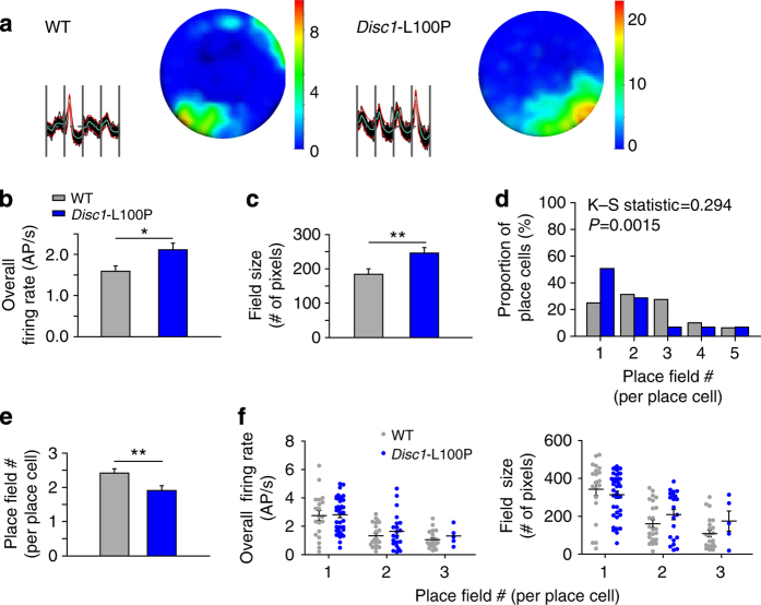
Results: The spatial specificity and stability of Disc1-L100P place cells were similar to wild-type place cells. Importantly, however, Disc1-L100P place cells exhibited a higher propensity to increase their firing rate in a single, large location of the environment, rather than multiple smaller locations, indicating a generalization in their spatial selectivity. Alterations in the signaling and numbers of CA1 putative inhibitory interneurons and decreased hippocampal theta (5-12 Hz) power were also identified in the Disc1-L100P mice.

Conclusions: The generalized spatial selectivity of Disc1-L100P place cells suggests a simplification of the ensemble place codes that encode individual locations and subserve spatial working memory. Moreover, these results suggest that deficient working memory in schizophrenia results from an impaired ability to uniquely code the individual components of a memory sequence.


Conflict of interest statement

The authors declare no conflict of interest.

Figures

Figure 1
Figure 1
CA1 place cells from mice with a L100P mutation in the disrupted-in-schizophrenia-1 gene (Disc1-L100P) exhibit intact spatial stability. (a) Schematic of cue rotation protocol and exemplar place field rotations from a wild-type (WT) and Disc1-L100P place cell. Color-coded firing rate maps are shown with corresponding frequency scaled to peak (red). (b) Intact coupling of place cell activity to cue between sessions 1 and 2 (upper) and sessions 1 and 3 (lower). Y-axis depicts the similarity score of place fields between sessions 1 and 2 at 270° (i.e., 90° counterclockwise upper graph) or sessions 1 and 3 at 0° (lower graph). N=20 WT and 37 Disc1-L100P place cells. (c) Intact place field coherence and information content scores in Disc1-L100P place cells. Plot whiskers denote minimum and maximum values. The top and bottom of boxes denote the first and third quartile, respectively. Mann–Whitney U-test. N=80 WT place cells and 73 Disc1-L100P place cells. CCW, counterclockwise; CW, clockwise; info. con. score, information content score.
Figure 2
Figure 2
Disc1-L100P mice exhibit fewer place fields per place cell. (a) Tetrode recordings and place fields from a wild-type (WT) and Disc1-L100P mouse. Vertical sections on tetrode traces distinguish recordings from individual tetrode wires. Note the difference in the number of fields and their size between the WT and Disc1-L100P place cell. (b) Disc1-L100P place cells show higher overall-firing rates. Data are represented as mean±s.e.m. Mann–Whitney U-test. (c) Disc1-L100P place cells exhibit larger and (d,e) fewer place fields. Two-sample Kolmogorov–Smirnov (K–S) test (d) and Mann–Whitney U-test (e), latter data represented as mean±s.e.m. (f) Comparison between place cells displaying equivalent place field numbers identified no differences between Disc1-L100P and WT place cells in average firing rate (left) or field size (right). N=80 WT and 73 Disc1-L100P place cells. *P<0.05, **P<0.01. AP, action potential.
Figure 3
Figure 3
Disc1-L100P mice exhibit aberrant inhibitory interneuron activity and deficits in CA1 parvalbumin-positive interneuron numbers. (a) Disc1-L100P place cells show lower signal-to-noise ratios, when compared with wild-type (WT) place cells, consistent with the elevated numbers of active interneurons recorded from Disc1-L100P mice (see Results for details). Plot whiskers denote minimum and maximum values. The top and bottom of boxes denote the first and third quartile, respectively. Mann–Whitney U-test. N=80 WT and 73 Disc1-L100P place cells. (b) Superimposed action potential traces recorded from a putative interneuron in a WT and a Disc1-L100P mouse. (c) Disc1-L100P putative interneurons exhibit steeper action potential rising slope (upper) and unaffected falling slopes (lower). Mann–Whitney U-test. N=19 WT and 38 Disc1-L100P putative interneurons. (d) Power spectrums from a WT and Disc1-L100P mouse. Theta band (5–12 Hz) is highlighted in red. (e) Disc1-L100P mice show reduced theta power. Data represented as mean±s.e.m. Kruskal–Wallis one-way analysis of variance (ANOVA) on ranks. N=6 WT and 6 Disc1-L100P recordings. (f) Example confocal images of anti-parvalbumin and anti-GAD67 fluorescence from a WT (upper) and Disc1-L100P (lower) central hippocampal section. Arrows point to some of the cells that stained positive for both parvalbumin and GAD67. Bar = 500 μm. (g) Disc1-L100P mice possess similar numbers of GAD67-positive cells in the central hippocampus. Plotted data show mean±s.e.m. two-way ANOVA. N=3 WT and 3 Disc1-L100P mice. (h) Disc1-L100P mice exhibit alterations in parvalbumin-positive interneuron numbers. Relative to WT mice, Disc1-L100P mice exhibit fewer parvalbumin-positive interneurons in the central CA1 (upper), more parvalbumin-positive interneurons in the rostral hippocampus (lower left), and similar levels of parvalbumin-positive interneurons in the caudal hippocampus (lower right). Plotted data show mean±s.e.m. two-way ANOVA. Data represents average of 9 to 23 sections from N=3 WT and 3 Disc1-L100P mice. *P<0.05, ***P<0.001. A/D, analog-to-digital; Disc1-L100P, disrupted-in-schizophrenia-1 gene; RMS, root mean square.

References

    1. Keefe RS, Roitman SE, Harvey PD, Blum CS, DuPre RL, Prieto DM, et al. A pen-and-paper human analogue of a monkey prefrontal cortex activation task: spatial working memory in patients with schizophrenia. Schizophr Res. 1995;17:25–33. doi: 10.1016/0920-9964(95)00027-J. - DOI - PubMed
    1. Park S, Holzman PS. Schizophrenics show spatial working memory deficits. Arch Gen Psychiatry. 1992;49:975–982. doi: 10.1001/archpsyc.1992.01820120063009. - DOI - PubMed
    1. Park S, Puschel J, Sauter BH, Rentsch M, Hell D. Spatial working memory deficits and clinical symptoms in schizophrenia: a 4-month follow-up study. Biol Psychiatry. 1999;46:392–400. doi: 10.1016/S0006-3223(98)00370-9. - DOI - PubMed
    1. Clapcote SJ, Lipina TV, Millar JK, Mackie S, Christie S, Ogawa F, et al. Behavioral phenotypes of Disc1 missense mutations in mice. Neuron. 2007;54:387–402. doi: 10.1016/j.neuron.2007.04.015. - DOI - PubMed
    1. Kvajo M, McKellar H, Arguello PA, Drew LJ, Moore H, MacDermott AB, et al. A mutation in mouse Disc1 that models a schizophrenia risk allele leads to specific alterations in neuronal architecture and cognition. Proc Natl Acad Sci USA. 2008;105:7076–7081. doi: 10.1073/pnas.0802615105. - DOI - PMC - PubMed