. 2016 Jun 23;7(6):e2271.
doi: 10.1038/cddis.2016.173.

Mitofusin-mediated ER stress triggers neurodegeneration in pink1/parkin models of Parkinson's disease

Affiliations

Mitofusin-mediated ER stress triggers neurodegeneration in pink1/parkin models of Parkinson's disease

I Celardo et al. Cell Death Dis. .

Abstract

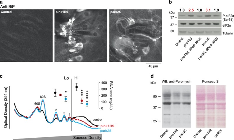
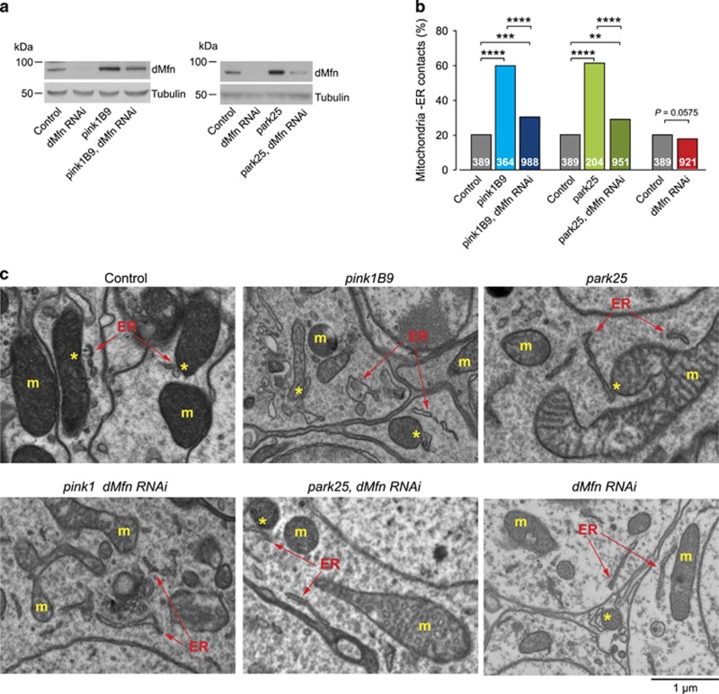
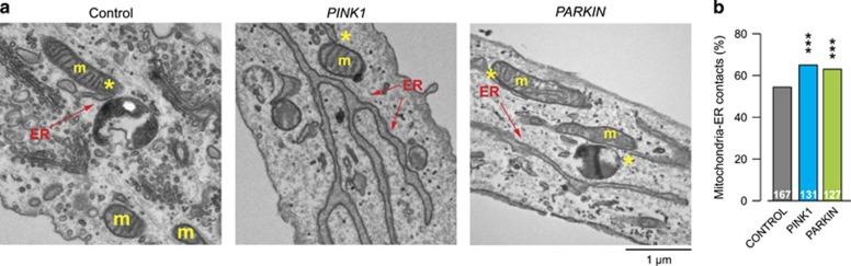
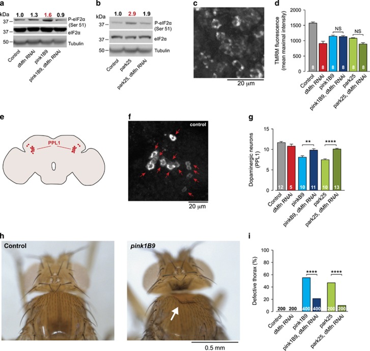
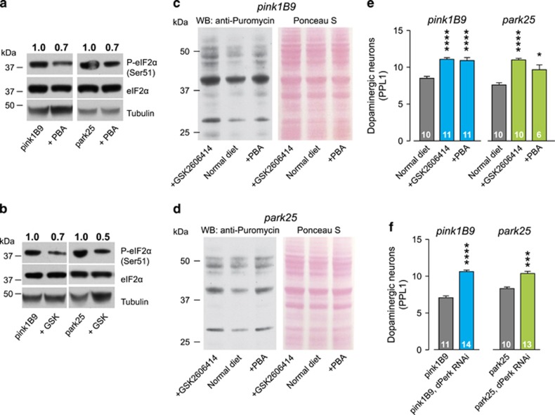
Mutations in PINK1 and PARKIN cause early-onset Parkinson's disease (PD), thought to be due to mitochondrial toxicity. Here, we show that in Drosophila pink1 and parkin mutants, defective mitochondria also give rise to endoplasmic reticulum (ER) stress signalling, specifically to the activation of the protein kinase R-like endoplasmic reticulum kinase (PERK) branch of the unfolded protein response (UPR). We show that enhanced ER stress signalling in pink1 and parkin mutants is mediated by mitofusin bridges, which occur between defective mitochondria and the ER. Reducing mitofusin contacts with the ER is neuroprotective, through suppression of PERK signalling, while mitochondrial dysfunction remains unchanged. Further, both genetic inhibition of dPerk-dependent ER stress signalling and pharmacological inhibition using the PERK inhibitor GSK2606414 were neuroprotective in both pink1 and parkin mutants. We conclude that activation of ER stress by defective mitochondria is neurotoxic in pink1 and parkin flies and that the reduction of this signalling is neuroprotective, independently of defective mitochondria. A video abstract for this article is available online in the supplementary information.


Figures

Figure 1
Figure 1
Activation of phospho-eIF2α signalling and attenuation of translation in pink1 and parkin mutant flies. (a) Increased levels of BiP in the body wall muscle of pink1 and parkin mutant larvae. Representative confocal images with the indicated genotype stained with α-BiP antibody are shown. (b) Increased levels of phospho-eIF2α in pink1 and parkin mutant flies are reduced by knockdown of dPerk. Whole-fly lysates were analysed using the indicated antibodies. Ratios of signal intensity between phospho and total-eIF2α are shown at the top. (c) Polysomal distribution of mRNAs of young adult male flies showing individual ribosomal subunits and the polysome peaks. RNA concentrations were measured from the low (Lo) and high (Hi) translation fractions (mean±S.D., asterisks, one-way ANOVA with Dunnett's multiple comparison test; n=4). (d) Reduced puromycin incorporation in nascent proteins in pink1 and parkin mutant flies. Whole-fly lysates were analysed with an anti-puromycin antibody and equivalent protein loading was assessed by Ponceau S staining of the membranes. Genotypes for (b) Control: daGAL4; pink1B9: pink1B9,daGAL4; park25: park25/park25,daGAL4. RNAi dPerk was driven by daGAL4. (a, c and d) Control: w1118
Figure 2
Figure 2
dMfn mediates the recruitment of defective mitochondria to ER contact points in pink1 or parkin mutants. (a) The RNAi-mediated suppression of dMfn reduces dMfn protein levels in pink1 and parkin mutants. Whole-fly lysates were analysed using the indicated antibodies. (b and c) Quantification of mitochondria–ER contacts in adult Drosophila brains (asterisks, chi-square two-tailed, 95% confidence intervals) (b), and representative electron microscopy images (c). Yellow asterisks show mitochondria in contact with ER (arrows). ER, endoplasmic reticulum; m, mitochondria. Genotypes for (a) Control: daGAL4; pink1B9: pink1B9,daGAL4; park25: park25/park25,daGAL4. RNAi dMfn was driven by daGAL4. (b and c) Control: elavGAL4; pink1B9: pink1B9,elavGAL4; park25: park25/park25,elavGAL4. RNAi dMfn was driven by elavGAL4
Figure 3
Figure 3
Increase in mitochondria–ER contact points in PINK1 or PARKIN mutant fibroblasts. (a and b) Quantification of mitochondria–ER contacts in human fibroblasts (asterisks, chi-square two-tailed, 95% confidence intervals) (b), and representative electron microscopy images (a). Yellow asterisks show mitochondria in contact with ER (arrows). ER, endoplasmic reticulum; m, mitochondria. PINK1: c.261_276del16; p.T90LfsX12 (homozygous); PARKIN: deletion of exons 3 and 4 (homozygous)
Figure 4
Figure 4
Suppressing dMfn in pink1 or parkin mutants attenuates eIF2α signalling and blocks neurodegeneration. (a and b) Decreased levels of phospho-eIF2α upon RNAi-mediated suppression of dMfn in pink1 (a) and parkin (b) mutant flies. Whole-fly lysates were analysed using the indicated antibodies. Ratios of signal intensity between phospho and total-eIF2α are shown at the top. (c and d) RNAi-mediated suppression of dMfn does not prevent the loss of Δψm in pink1 or parkin mutants. Representative confocal image of a whole mounted control brain showing neurons loaded with TMRM (c). Quantification of Δψm in the brains of the indicated genotypes (d) (mean±S.E.M.; asterisks, one-way ANOVA with Bonferroni's multiple comparison test). (eg) The RNAi-mediated suppression of dMfn rescues the loss of dopaminergic neurons in the PPL1 cluster of pink1 and parkin mutant flies. Schematic diagram of a fly brain in sagittal orientation indicating the PPL1 cluster of dopaminergic neurons in red (e). Anti-TH staining showing cell bodies of PPL1 neurons in a representative control brain (f) and quantification of the PPL1 cluster neurons (g) (mean±S.E.M.; asterisks, one-way ANOVA with Bonferroni's multiple comparison test). (h and i) Suppression of the thoracic defects of pink1 and parkin mutants by RNAi-mediated suppression of dMfn. Representative images of normal and defective thorax in pink1 mutants, the arrow points to a thoracic defect (h). Quantification of the thoracic defects (i) in the indicated genotypes (asterisks, chi-square two-tailed, 95% confidence intervals). Genotypes for (a, b, i) Control: daGAL4; pink1B9: pink1B9,daGAL4; park25: park25/park25,daGAL4. RNAi dMfn was driven by daGAL4. (c, d, f, g) Control: elavGAL4; pink1B9: pink1B9,elavGAL4; park25: park25/park25,elavGAL4. RNAi dMfn was driven by elavGAL4. (h) Control: w1118
Figure 5
Figure 5
Inhibiting PERK/eIF2α signalling in pink1 and parkin mutants prevents neurodegeneration. (a and b) The chemical chaperone PBA (a) and the PERK inhibitor GSK2606414 (b) decrease the levels of phospho-eIF2α in pink1 and parkin mutant flies. Whole-fly lysates were analysed using the indicated antibodies. Ratios of signal intensity between phospho and total-eIF2α are shown at the top. (c and d) Recovery of puromycin incorporation in nascent proteins in pink1 (c) and parkin (d) mutant flies upon dietary supplementation with PBA or GSK2606414. Whole-fly lysates were analysed with an anti-puromycin antibody and equivalent protein loading was assessed by Ponceau S staining of the membranes. (e) The chemical chaperone PBA and the PERK inhibitor GSK2606414 rescue the loss of dopaminergic neurons in the PPL1 cluster of pink1 and parkin mutant flies (mean±S.E.M.; asterisks, one-way ANOVA with Bonferroni's multiple comparison test). (f) The RNAi-mediated suppression of dPerk rescues the loss of dopaminergic neurons in the PPL1 cluster of pink1 and parkin mutant flies (mean±S.E.M.; asterisks, one-way ANOVA with Bonferroni's multiple comparison test). Genotypes for (f) pink1B9: pink1B9,elavGAL4; park25: park25/park25,elavGAL4. RNAi dPerk was driven by elavGAL4

References

    1. Halliday M, Mallucci GR. Targeting the unfolded protein response in neurodegeneration: a new approach to therapy. Neuropharmacology 2014; 76 Pt A: 169–174. - PubMed
    1. Scheper W, Hoozemans JJ. The unfolded protein response in neurodegenerative diseases: a neuropathological perspective. Acta Neuropathol 2015; 130: 315–331. - PMC - PubMed
    1. Moreno JA, Radford H, Peretti D, Steinert JR, Verity N, Martin MG et al. Sustained translational repression by eIF2alpha-P mediates prion neurodegeneration. Nature 2012; 485: 507–511. - PMC - PubMed
    1. Radford H, Moreno JA, Verity N, Halliday M, Mallucci GR. PERK inhibition prevents tau-mediated neurodegeneration in a mouse model of frontotemporal dementia. Acta Neuropathol 2015; 130: 633–642. - PMC - PubMed
    1. Moreno JA, Halliday M, Molloy C, Radford H, Verity N, Axten JM et al. Oral treatment targeting the unfolded protein response prevents neurodegeneration and clinical disease in prion-infected mice. Sci Transl Med 2013; 5: 206ra138. - PubMed

MeSH terms