. 2016 Jun 23;7(6):e2275.
doi: 10.1038/cddis.2016.179.

Junctophilin 3 expresses in pancreatic beta cells and is required for glucose-stimulated insulin secretion

Affiliations

Junctophilin 3 expresses in pancreatic beta cells and is required for glucose-stimulated insulin secretion

L Li et al. Cell Death Dis. .

Abstract

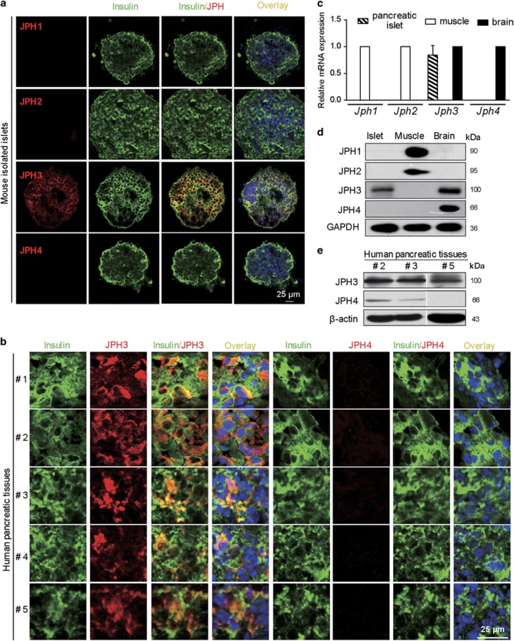
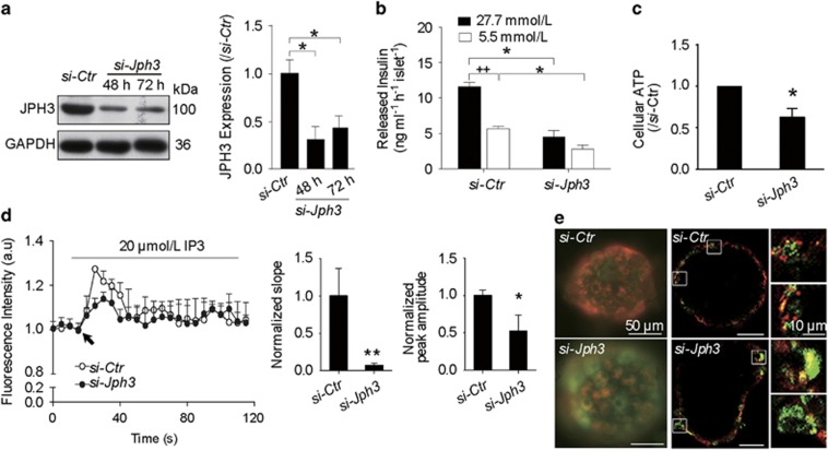
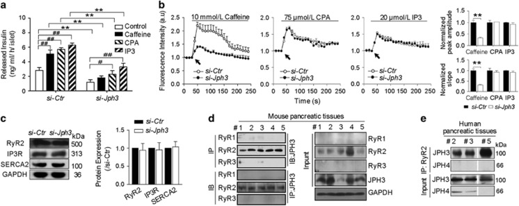
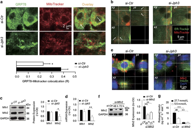
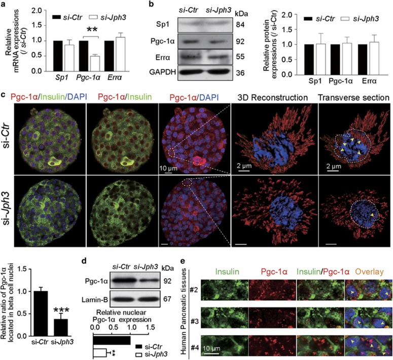
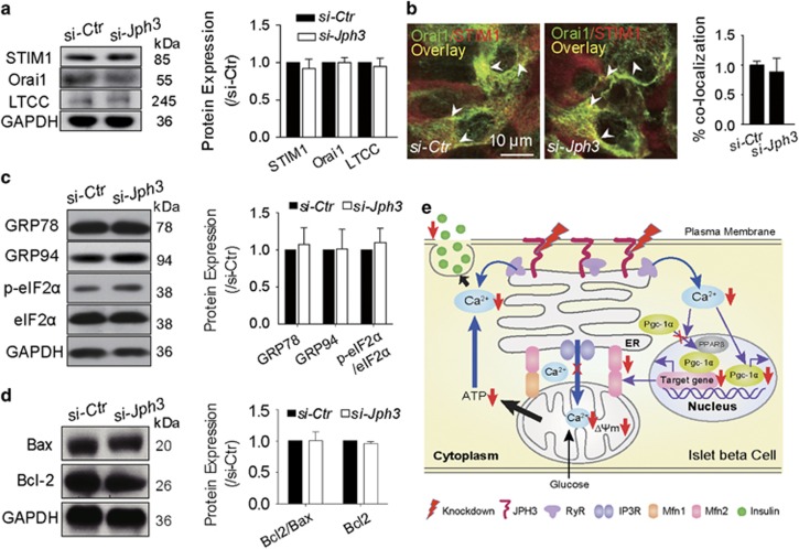
It is well accepted that junctophilin (JPHs) isoforms act as a physical bridge linking plasma membrane and endoplasmic reticulum (ER) for channel crosstalk in excitable cells. Our purpose is to investigate whether JPHs are involved in the proper communication between Ca(2+) influx and subsequent Ca(2+) amplification in pancreatic beta cells, thereby participating in regulating insulin secretion. The expression of JPH isoforms was examined in human and mouse pancreatic tissues, and JPH3 expression was found in both the beta cells. In mice, knockdown of Jph3 (si-Jph3) in islets decreased glucose-stimulated insulin secretion (GSIS) accompanied by mitochondrial function impairment. Si-Jph3 lowered the insulin secretory response to Ca(2+) signaling in the presence of glucose, and reduced [Ca(2+)]c transient amplitude triggered by caffeine. Si-Jph3 also attenuated mitofusin 2 expression, thereby disturbing the spatial organization of ER-mitochondria contact in islets. These results suggest that the regulation of GSIS by the KATP channel-independent pathways is partly impaired due to decrease of JPH3 expression in mouse islets. JPH3 also binds to type 2 ryanodine receptors (RyR2) in mouse and human pancreatic tissues, which might contribute to Ca(2+) release amplification in GSIS. This study demonstrates some previously unrecognized findings in pancreatic tissues: (1) JPH3 expresses in mouse and human beta cells; (2) si-Jph3 in mouse primary islets impairs GSIS in vitro; (3) impairment in GSIS in si-Jph3 islets is due to changes in RyR2-[Ca(2+)]c transient amplitude and ER-mitochondria contact.


Figures

Figure 1
Figure 1
JPHs expression identification. (a) JPH3 in mouse islets. (b) JPH3 in human pancreatic tissues. (c and d) Jph3 mRNA and JPH3 protein levels in mouse islets, mouse skeletal muscle and brain tissues as positive control. (e) JPH3 and JPH4 in human pancreatic tissues. Bar=25 μm. See also Supplementary Table 1 for patient donor information
Figure 2
Figure 2
GSIS and mitochondrial function in si-Jph3 islets. (a) Jph3 silencing efficiency. (b) GSIS in response to high and low levels of glucose. (c) Intracellular ATP generation in the presence of glucose. (d) IP3-evoked [Ca2+]m transients amplitude in permeabilized islets. Data are means±S.D. n=3 mice per group, with the minimum 30 islets for GSIS or 20 islets for ATP per mouse. *P<0.05, **P<0.01 versus si-Ctr. ++P<0.01 versus 27.7 mmol/L glucose. (e) Global ΔΨm in islets. Bar=10 μm
Figure 3
Figure 3
Insulin secretory response to Ca2+ signaling and [Ca2+]c transient amplitude in si-Jph3 islets. (a) Insulin secretory response to Ca2+ signaling in the presence of glucose, and (b) [Ca2+]c transient amplitude in response to chemicals. n=3 mice per group, with the minimum 30 islets per mouse for insulin secretion. (c) Expression of Ca2+ releasing proteins of ER (n=3). (d and e) Co-immunoprecipitation showed JPH3 binds to RyR2 in mouse (n=5) and human (n=3) pancreatic tissues. Data are means±S.D. *P<0.05, **P< 0.01 versus si-Ctr. #P<0.05, ##P<0.01 versus control in the same group
Figure 4
Figure 4
ER–mitochondria contact and Mfn2-dependent GSIS in si-Jph3 islets. (a) The colocalization of ER and mitochondria in islets. (b) 3D confocal images of ER–mitochondria contact. (c and d) Mfn2 protein and Mfn2 mRNA expression (n=3). (e) 3D confocal images of Mfn2 and insulin. (f) Mfn2 silencing efficiency (n=3). (g) GSIS in si-Mfn2 islets, n=3 mice per group, with the minimum 30 islets per mouse. Data are means±S.D. *P<0.05, **P<0.01 versus si-Ctr. ##P<0.01 versus 27.7 mmol/L glucose
Figure 5
Figure 5
Pgc-1α in islets. (a and b) Expressions of Pgc-1α mRNA and Pgc-1α protein (n=3). (c) Confocal laser scanning exhibited the colocalization of Pgc-1α with insulin in si-Ctr and si-Jph3 islets. Three-dimensional reconstruction and transverse section of a beta cell further displayed the localization of Pgc-1α within a nucleus (yellow arrowhead). Ratio of Pgc-1α existed in nuclei were showed in bar chart (n=4). (d) Expression of nuclear Pgc-1α in si-Ctr and si-Jph3 islets (n=4). (e) Pgc-1α existed in nuclei of human beta cells. Data are means±S.D. *P<0.05, **P<0.01 versus si-Ctr
Figure 6
Figure 6
Ca2+ influx and ER stress or Bcl-2/Bax-related proteins in si-Jph3 islets. Unchanged expression of Ca2+ influx-related proteins (a), colocalization areas (b), ER stress protein (c) Bcl-2/Bax ratio (d). n=3. (e) Schematic representation of the mechanism of JPH3-dependent GSIS in mouse beta cells

Similar articles

Cited by

References

    1. Santulli G, Pagano G, Sardu C, Xie W, Reiken S, D'Ascia SL et al. Calcium release channel RyR2 regulates insulin release and glucose homeostasis. J Clin Invest 2015; 125: 4316. - PMC - PubMed
    1. Allagnat F, Klee P, Cardozo AK, Meda P, Haefliger JA. Connexin36 contributes to INS-1E cells survival through modulation of cytokine-induced oxidative stress, ER stress and AMPK activity. Cell Death Differ 2013; 20: 1742–1752. - PMC - PubMed
    1. Ravier MA, Daro D, Roma LP, Jonas JC, Cheng-Xue R, Schuit FC et al. Mechanisms of control of the free Ca2+ concentration in the endoplasmic reticulum of mouse pancreatic beta-cells: interplay with cell metabolism and [Ca2+]c and role of SERCA2b and SERCA3. Diabetes 2011; 60: 2533–2545. - PMC - PubMed
    1. Luciani DS, Gwiazda KS, Yang TL, Kalynyak TB, Bychkivska Y, Frey MH et al. Roles of IP3R and RyR Ca2+ channels in endoplasmic reticulum stress and beta-cell death. Diabetes 2009; 58: 422–432. - PMC - PubMed
    1. Demozay D, Tsunekawa S, Briaud I, Shah R, Rhodes CJ. Specific glucose-induced control of insulin receptor substrate-2 expression is mediated via Ca2+-dependent calcineurin/NFAT signaling in primary pancreatic islet beta-cells. Diabetes 2011; 60: 2892–2902. - PMC - PubMed

MeSH terms