. 2016 Jun 23;10(6):e0004746.
doi: 10.1371/journal.pntd.0004746. eCollection 2016 Jun.

An mRNA Vaccine Encoding Rabies Virus Glycoprotein Induces Protection against Lethal Infection in Mice and Correlates of Protection in Adult and Newborn Pigs

Affiliations

An mRNA Vaccine Encoding Rabies Virus Glycoprotein Induces Protection against Lethal Infection in Mice and Correlates of Protection in Adult and Newborn Pigs

Margit Schnee et al. PLoS Negl Trop Dis. .

Abstract

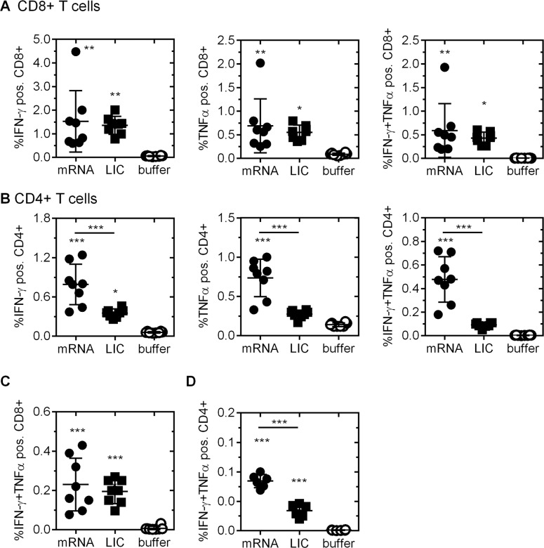
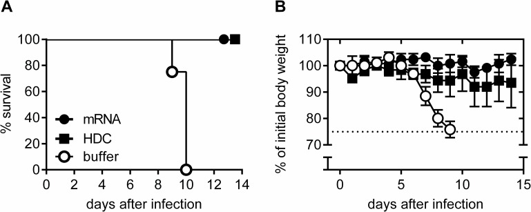
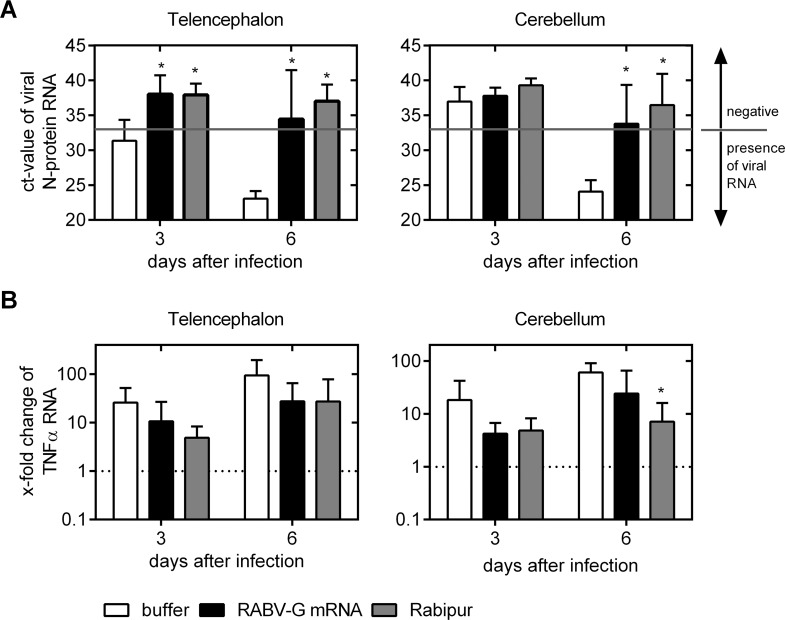
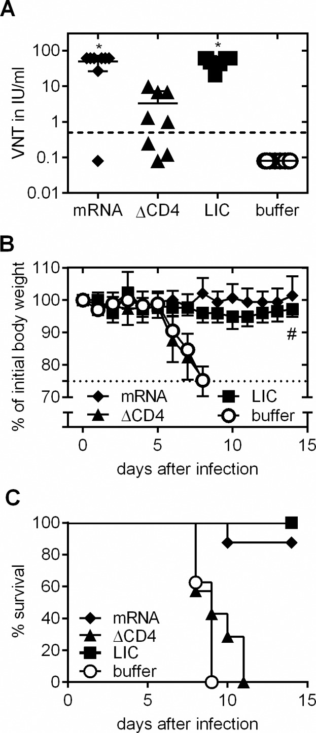
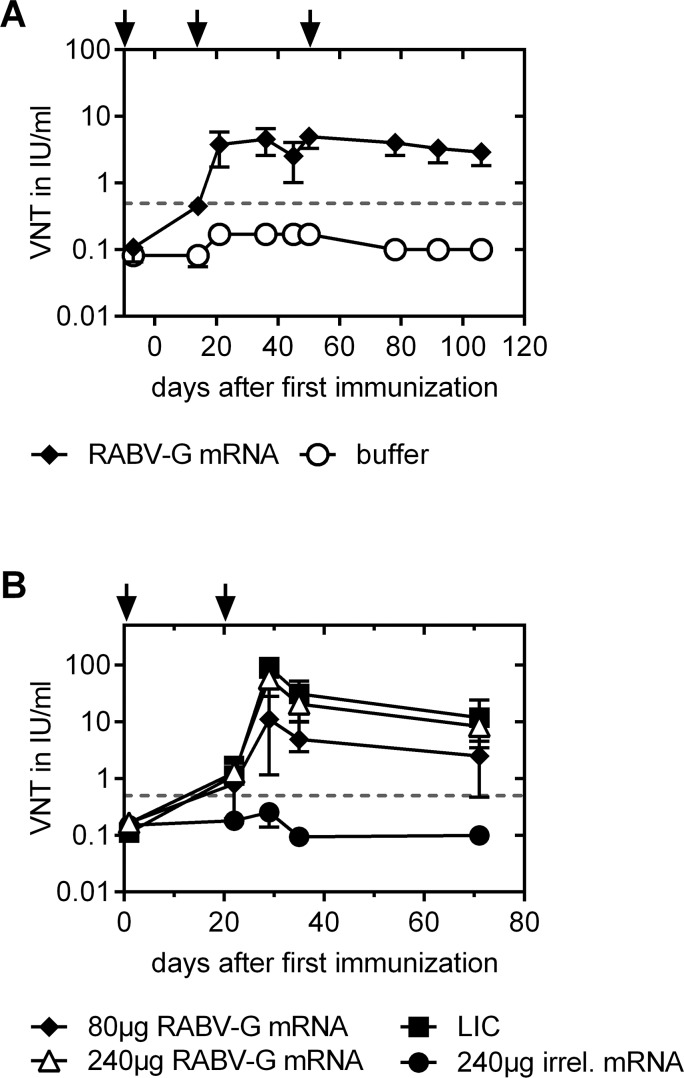
Rabies is a zoonotic infectious disease of the central nervous system (CNS). In unvaccinated or untreated subjects, rabies virus infection causes severe neurological symptoms and is invariably fatal. Despite the long-standing existence of effective vaccines, vaccine availability remains insufficient, with high numbers of fatal infections mostly in developing countries. Nucleic acid based vaccines have proven convincingly as a new technology for the fast development of vaccines against newly emerging pathogens, diseases where no vaccine exists or for replacing already existing vaccines. We used an optimized non-replicating rabies virus glycoprotein (RABV-G) encoding messenger RNA (mRNA) to induce potent neutralizing antibodies (VN titers) in mice and domestic pigs. Functional antibody titers were followed in mice for up to one year and titers remained stable for the entire observation period in all dose groups. T cell analysis revealed the induction of both, specific CD4+ as well as CD8+ T cells by RABV-G mRNA, with the induced CD4+ T cells being higher than those induced by a licensed vaccine. Notably, RABV-G mRNA vaccinated mice were protected against lethal intracerebral challenge infection. Inhibition of viral replication by vaccination was verified by qRT-PCR. Furthermore, we demonstrate that CD4+ T cells are crucial for the generation of neutralizing antibodies. In domestic pigs we were able to induce VN titers that correlate with protection in adult and newborn pigs. This study demonstrates the feasibility of a non-replicating mRNA rabies vaccine in small and large animals and highlights the promises of mRNA vaccines for the prevention of infectious diseases.


Conflict of interest statement

I have read the journal's policy and the authors of this manuscript have the following competing interests: MS, DV, BP, PB and TK are or were employees of CureVac. Authors MS, TK, BP and LS are named as inventors on a patent application for a rabies vaccine filed by CureVac.

Figures

Fig 1
Fig 1. Antigen-specific immune response in mice.
(A) Virus-neutralizing (VN) antibody titers in the serum of female BALB/c mice were measured using Fluorescent Antibody Virus Neutralization (FAVN) test. Animals were vaccinated twice with 80 μg RABV-G mRNA or buffer at day 0 and 21. 35 days after the first vaccination, serum was collected. The dotted line at 0.5 IU/ml is considered as a correlate of protection. (B) Splenocytes from the same mice were isolated for the analysis of antigen specific T cells via ELISPOT analysis at day 35 after the first immunization. (C, D) VN titers dose-response profile in female (C) C57BL/6 and (D) BALB/c mice after RABV-G mRNA vaccination compared to licensed vaccines. Animals were vaccinated with RABV-G mRNA, licensed vaccines (HDC or Rabipur (LIC)) or buffer on study days 0 and 21. RABV-G mRNA was applied intradermally (i.d.) at doses of 80 μg, 40 μg, 20 μg, 10 μg, 5 μg, 2.5 μg or 1.25 μg. For positive control, 100 μl (0.1 human dose) of Rabipur and HDC were administered intramuscularly (i.m.). Presence of rabies-specific VN titers in sera of vaccinated and control mice were analyzed 2 weeks after immunization using the FAVN test. In both graphs the mean and standard deviation (SD) is plotted and significance compared to buffer treated mice was analysed using one-way ANOVA Dunnett’s multiple comparisons test (* p<0.01). (E) Kinetic of virus-neutralizing antibodies in BALB/c mice from (D). Immunization points are indicated by arrows. Mean an SD is presented (n = 8/group).
Fig 2
Fig 2. Induction of rabies virus specific T cells upon immunization of female BALB/c mice with RABV-G mRNA.
Mice were vaccinated twice (days 0 and 21) with 80 μg RABV-G mRNA, 0.1 human dose Rabipur (LIC) or buffer. Activated, antigen specific (A) CD8+ T cells and (B) CD4+ T cells were analyzed by intracellular staining of IFN-γ alone (left panel), TNFα alone (middle panel) or IFN-γ and TNFα (right panel) followed by flow cytometry analysis. Isolation of splenocytes was done 6 days after the second immunization. (C+D) Analysis of long-lasting T cell immunoreactivity. Mice were treated with 80 μg RABV-G mRNA, 0.1 human dose Rabipur (LIC) or injection buffer alone at days 0 and 21. Ten weeks after boost, animals were sacrificed and IFN-γ and TNFα double positive antigen-specific (C) CD8+ and (D) CD4+ T cells were analysed by flow cytometry (n = 8/group). Mean and SD (n = 8/group) is presented. Statistical significance was tested with one-way ANOVA test using Tukey’s multiple comparisons test compared to buffer group or as indicated (* p≤0.05; ** p≤0.01; *** p≤0.0001).
Fig 3
Fig 3. Protective capacity of mRNA vaccine against lethal intracerebal (i.c.) rabies challenge infection.
Female BALB/c mice were vaccinated twice (days 0 and 21) with 80 μg RABV-G mRNA, 0.1 human dose HDC or injection buffer alone as negative control. Six weeks after the second immunization, mice were infected i.c. with rabies virus. (A) Survival of challenged mice is illustrated by a Kaplan-Meyer-analysis. (B) Body weight of challenged mice was monitored daily. As soon as animals lost 25% of initial body weight (dotted line), animals were sacrificed for reasons of humane endpoint criteria (n = 5 /group). Mean and SD is presented.
Fig 4
Fig 4. qRT-PCR analysis of viral N-protein encoding RNA and cellular TNFα mRNA transcript levels in brains 3 and 6 days after i.c. rabies challenge infection.
Female BALB/c mice were injected with buffer alone (white bar), 80 μg RABV-G mRNA (black bar) or 0.1 human dose Rabipur (grey bar), on study days 0 and 21. 5 weeks after second immunization, mice were i.c. infected (into the telencephalon) with rabies virus. 3 and 6 days thereafter, mice were sacrificed. Perfused brains were divided into telencephalon (left panel) and cerebellum (right panel) and tested by qRT-PCR. (A) Detection of viral N-protein RNA. Ct-values are illustrated with a N-protein RNA detection limit of 33 cycles (grey line). Ct-values below the detection limit are positive for virus RNA as indicated in the figure. Bars represent the mean and SD (n = 4/group). Significance between the buffer control group (white bar) and immunized groups was calculated using the one-way ANOVA Tukey’s multiple comparison test (* p<0.05). (B) TNFα mRNA levels in the telencephalon (left panel) and cerebellum (right panel). RNA of brains from uninfected animals served as control. mRNA values of controls were defined as “1” (dotted line). Fold changes were calculated and bars represent the mean and SD (n = 4/group). Statistical significance to uninfected controls was tested with one-way ANOVA Tukey’s multiple comparison test with a significance level of 5% (*).
Fig 5
Fig 5. CD4+ T cell depletion during immunization phase followed by challenge infection.
Female BALB/c mice were vaccinated with RABV-G mRNA, Rabipur (LIC) or injection buffer alone on days 0 and 21. The CD4+ T cell population was specifically depleted in one mRNA-receiving group by injection of anti-CD4 antibody on days -1, 1, 20 and 22. (A) Evaluation of rabies virus neutralizing antibodies in sera of control, vaccinated or vaccinated and CD4-depleted mice 4 weeks after the second immunization using the FAVN test (n = 8/group). Mean and SD is presented. Significance between the buffer control group and immunized groups was calculated using the one-way ANOVA Tukey’s multiple comparison test (* p<0.05). (B, C) Mice were challenged with a lethal dose of rabies virus and (B) body weight was monitored daily. A loss of 25% of initial body weight was defined as humane endpoint. Mean and SD is presented. (C) Survival of challenged mice. # note: the sacrificed mouse in the RABV-G mRNA (black diamond) group was excluded from the body weight analysis to demonstrate the healthy state of the remaining animals. This mouse was also negative for rabies specific antibodies (see Fig 5A).
Fig 6
Fig 6. Seroconversion profile after RABV-G mRNA and Rabipur (LIC) vaccination in domestic pigs.
Immunization points are indicated by arrows. (A) Adult pigs received intradermal immunizations on days 0, 14, 49 with either 80 μg RABV-G mRNA or injection buffer alone. At different time points after immunization, sera were tested for rabies-specific neutralizing antibodies using FAVN testing (n = 6 pigs/group). (B) Newborn piglets were immunized in their first week of life and 3 weeks later with 80 μg or 240 μg RABV-G mRNA (i.d.) or a full human dose of Rabipur (LIC; i.m.). As a negative control, animals were immunized with 240 μg of an irrelevant mRNA. At different time points blood samples were taken and sera were tested for virus-neutralizing antibodies using the FAVN test (n = 5 or n = 6 (240 μg) piglets/group). In both graphs mean and SD is illustrated.

References

    1. Hemachudha T, Ugolini G, Wacharapluesadee S, Sungkarat W, Shuangshoti S, et al. (2013) Human rabies: neuropathogenesis, diagnosis, and management. Lancet Neurology 12: 498–513. 10.1016/S1474-4422(13)70038-3 - DOI - PubMed
    1. Pathak S, Horton DL, Lucas S, Brown D, Quaderi S, et al. (2014) Diagnosis, management and post-mortem findings of a human case of rabies imported into the United Kingdom from India: a case report. Virology Journal 11. - PMC - PubMed
    1. Ertl HCJ (2009) Novel vaccines to human rabies. PLoS neglected tropical diseases 3. - PMC - PubMed
    1. Wiktor TJ, György E, Schlumberger D, Sokol F, Koprowski H (1973) Antigenic properties of rabies virus components. Journal of Immunology (Baltimore, Md: 1950) 110: 269–276. - PubMed
    1. Cox JH, Dietzschold B, Schneider LG (1977) Rabies virus glycoprotein. II. Biological and serological characterization. Infection and Immunity 16: 754–759. - PMC - PubMed

Publication types

MeSH terms