. 2016 Jun 23;11(6):e0157786.
doi: 10.1371/journal.pone.0157786. eCollection 2016.

In Vitro Infection with Dengue Virus Induces Changes in the Structure and Function of the Mouse Brain Endothelium

Affiliations

In Vitro Infection with Dengue Virus Induces Changes in the Structure and Function of the Mouse Brain Endothelium

Myriam L Velandia-Romero et al. PLoS One. .

Abstract

Background: The neurological manifestations of dengue disease are occurring with greater frequency, and currently, no information is available regarding the reasons for this phenomenon. Some viruses infect and/or alter the function of endothelial organs, which results in changes in cellular function, including permeability of the blood-brain barrier (BBB), which allows the entry of infected cells or free viral particles into the nervous system.

Methods: In the present study, we standardized two in vitro models, a polarized monolayer of mouse brain endothelial cells (MBECs) and an organized co-culture containing MBECs and astrocytes. Using these cell models, we assessed whether DENV-4 or the neuro-adapted dengue virus (D4MB-6) variant infects cells or induces changes in the structure or function of the endothelial barrier.

Results: The results showed that MBECs, but not astrocytes, were susceptible to infection with both viruses, although the percentage of infected cells was higher when the neuro-adapted virus variant was used. In both culture systems, DENV infection changed the localization of the tight junction proteins Zonula occludens (ZO-1) and Claudin-1 (Cln1), and this process was associated with a decrease in transendothelial resistance, an increase in macromolecule permeability and an increase in the paracellular passing of free virus particles. MBEC infection led to transcriptional up-regulation of adhesion molecules (VCAM-1 and PECAM) and immune mediators (MCP-1 and TNF- α) that are associated with immune cell transmigration, mainly in D4MB-6-infected cells.

Conclusion: These results indicate that DENV infection in MBECs altered the structure and function of the BBB and activated the endothelium, affecting its transcellular and paracellular permeability and favoring the passage of viruses and the transmigration of immune cells. This phenomenon can be harnessed for neurotropic and neurovirulent strains to infect and induce alterations in the CNS.


Conflict of interest statement

Competing Interests: The authors have declared that no competing interests exist.

Figures

Fig 1
Fig 1. Phase contrast photomicrography of MBECs and astrocytes.
(A). Mouse brain endothelial cells were isolated and cultured for 30 days. At 15 days post seeding cells appear fusiform with different length cytoplasmic prolongations. At day 28, cells had polygonal morphology and monolayer acquired the cobblestone endothelial pattern. Bar correspond to 100 μm. To obtain astrocytes, mixed cell brain homogenates were cultured for 14 days and then shaken to detach non-related cells and to purify adhered astrocytes. Figure (left lower) shows the cell morphology at 8 days after purification by shaking, which displayed a typical star-shaped morphology with short cell prolongations. At 16 days post-seeding (right lower panel) cells displayed a morphology with short prolongations like the protoplasmic mature astrocytes. Bar = 20 μm. (B). Immunofluorescence detecting von Willebrand factor (vWF) in 99% of the MBECs. The tight junction proteins ZO-1 and Cln-1 were detected at 28 days post-seeding lining the plasma membrane, confirming the establishment of the barrier. Astrocytes showed 95% purity after staining with the specific markers GFAP and GLT-1. Their morphology was polygonal with many short prolongations and a small nucleus. Bar = 20 μm.
Fig 2
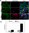
Fig 2. MBEC susceptibility to DENV infection.
(A). MBEC cultured on glass coverslips were inoculated for 48 h with mock suspension or infected with DENV-4 or D4MB-6 at an MOI:1 and stained for detecting viral envelope protein (red) and ZO-1 protein (green). Both viruses infected the MBECs and showed perinuclear localization of viral E protein (arrow). On the other hand, the typical plasma membrane localization pattern of ZO-1 was detected in the mock and DENV-4 infected cultures, while in the D4MB-6 infected cells was located mainly in the cytoplasm, in clusters or in a discontinuous pattern in the perimembrane region. Bar = 20 μm. (B). Infection percentages of MBEC after 24 or 48 h p.i. Infection proportion was significantly higher with D4MB-6 at 48 h p.i. (49%), with regard to 11% of the cells infected with DENV-4. Data are shown as the mean +/- SD of 3 independent cultures performed by duplicate.
Fig 3
Fig 3. Scheme barrier models and evaluation of TEER and permeability assay in each models.
(A). Endothelial barrier model scheme. Transwell inserts were used to establish the two barrier models. The first one (Monolayer model) consisted in MBEC cultured on the luminal side of the membrane (upper side) for four days until monolayer reaches TEER values between 1 to 1,5 kΩ. For establishing the second one barrier model (co-culture model), the glial cells were seed on the abluminal side inverting the insert for three days, then it was flipped to the right position before seeding the MBEC in the luminal side. (B). Transendothelial electrical resistance. MBEC in each barrier model were infected or treated with mock inoculum. Since 10 h p.i. there was a significant reduction in TEER in DENV-4 and D4MB-6 infected compared with mock-inoculated barrier models. This TEER loss was sustained up to 48 h p.i. (p<0.05, Kruskal-Wallis and Bonferroni tests), however, TEER changes were less drastic in MBEC-astrocytes co-culture with regard to monolayer barrier model. (C). Permeability assay. Using the same culture and infection protocols described above, dextran blue (DB) permeability assays were performed. DB was added in the insert’s upper chamber and at each time point; the lower chamber medium culture was collected to quantify the DB pass through by spectrophotometry. Since 10 to 48 h p.i. the infection with both virus strains induced a significant increase in lower chamber DB concentration coinciding with TEER loss. Barrier damage and DB quantified in the lower medium of MBEC-astrocytes co-culture were too low (between 0,2 to 1,2%) indicating a protective role of astrocytes. Data shown are mean of TEER or DB percentage from triplicates of two independent cultures and SD.
Fig 4
Fig 4. Immunostaining for ZO-1 in MBEC monolayer cultured on the inserts at 10 h p.i.
ZO-1 (in red) displayed the expected continuous membrane lining fluorescence pattern along the cell margins of each endothelial cell. At 10 h p.i. with DENV-4 or after mock treatment this fluorescence distribution did not change. By contrast, at 10 h p.i. MBEC infected with D4MB-6 showed slight changes in morphology (some cell detached) and ZO-1 redistribution from margin to cytoplasm, a finding correlated with the initial decrease in TEER values and dextran blue pass through the membrane. Bar = 20 μm.
Fig 5
Fig 5. ZO-1 protein redistribution at 24 and 48 h p.i., in DENV infected MBECs.
ZO-1 fluorescence pattern (green) in both barrier models infected with DENV-4 and D4MB-6 virus strains were evaluated one, and two days post infection. DENV-4 parental virus infection at 24 h p.i. did not affect the ZO-1 pattern in MBEC cells neither in monolayer barrier model nor in co-culture barrier model. On the contrary, neuroadapted dengue virus induced a ZO-1 re-localization from the cell margin to cytoplasm leaving a disrupted linear fluorescence pattern in the membrane. At 48 h p.i. both viruses induce cell detachment, however in the MBEC monolayer model was more severe with great cell loss and full ZO-1 rearrangement. Bar = 20 μm.
Fig 6
Fig 6. RNA quantitation of viruses, cell proteins, inflammatory mediators and adhesion molecules.
MBECs were infected with D4MB-6 and then processed to amplify a segment of dengue M protein gene, and cellular transcripts to ZO-1, TNF-α, MCP-1, PECAM, and VCAM using a quantitative RT-PCR. There were no significant changes in the amount of viral RNA and ZO-1 expression in the evaluated time points. TNF-α transcription was increased at 24 h p.i., while MCP-1 expression was increased at 10 h p.i. PECAM transcription increased at 2 and 24 h p.i. but not at 10 h p.i., while, VCAM expression was increased early (2 h p.i) and then gradually decreased over time. The data are shown as the relative expression obtained from duplicates of two independent experiments. The data were analyzed using the methods described in Pffafl et al [23] or Schefe et al, [24].
Fig 7
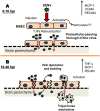
Fig 7. DENV transmigration model of passage through the BBB.
(A). After the virus inoculation, MBECs are activated and/or infected, inducing early TJP relocalization and the expression of MCP-1 and adhesion molecules (10 h p.i.). These changes allowed viral paracellular transport by which the particles passed through to abluminal side. Adhesion molecule expression also changed, bringing immune cells closer and favor rolling. (B). After viral replication in MBEC, transcellular virus transport occurs, and the integrity of the barrier is damaged, resulting in an increase in virus paracellular transport to the abluminal side. MBEC activation and the production of inflammatory mediators promote leukocyte adhesion and transmigration, which carries the virus through via a Trojan horse mechanism.

Similar articles

Cited by

References

    1. Abbott NJ, Dynamics of CNS Barriers: Evolution, Differentiation, and Modulation. Cell Mol Neurobiol. 2005, 25(1), 5–23. 10.1007/s10571-004-1374-y - DOI - PMC - PubMed
    1. Haseloff RF, Blasig IE, Bauer HC, Bauer H. In search of the astrocytic factor(s) modulating blood-brain barrier functions in brain capillary endothelial cells in vitro. Cell Mol Neurobiol. 2005, 1: 25–39. 10.1007/s10571-004-1375-x - DOI - PMC - PubMed
    1. Olsen AL Morrey JD, Smee DF, Sidwell RW. Correlation between breakdown of the blood–brain barrier and disease outcome of viral encephalitis in mice. Antiviral Res. 2007, 75, 104–112. 10.1016/j.antiviral.2006.11.013 - DOI - PMC - PubMed
    1. Spindler KR, Hsu TH. Viral disruption of the blood–brain barrier. Trends Microbiol. 2012, 20(6), 282–290. 10.1016/j.tim.2012.03.009 - DOI - PMC - PubMed
    1. Basu A, Chaturvedi UC. Vascular endothelium: the battlefield of dengue viruses. FEMS Immunol Med Microbiol. 2008, 53, 287–329. - PMC - PubMed

MeSH terms

Substances

LinkOut - more resources