. 2016 Jun 23:7:11974.
doi: 10.1038/ncomms11974.

Arginine demethylation is catalysed by a subset of JmjC histone lysine demethylases

Affiliations

Arginine demethylation is catalysed by a subset of JmjC histone lysine demethylases

Louise J Walport et al. Nat Commun. .

Abstract

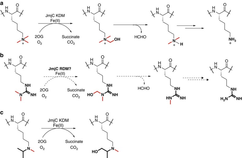
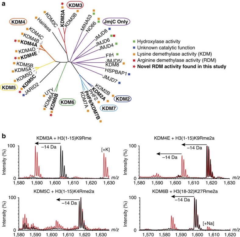
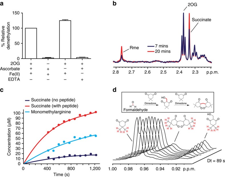
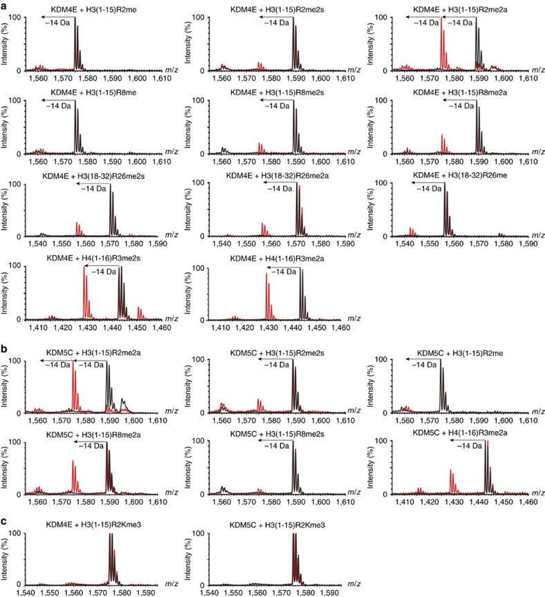
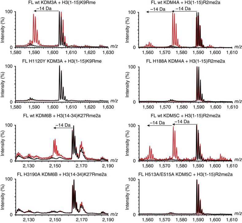
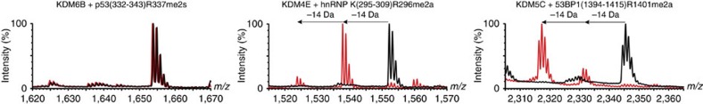
While the oxygen-dependent reversal of lysine N(ɛ)-methylation is well established, the existence of bona fide N(ω)-methylarginine demethylases (RDMs) is controversial. Lysine demethylation, as catalysed by two families of lysine demethylases (the flavin-dependent KDM1 enzymes and the 2-oxoglutarate- and oxygen-dependent JmjC KDMs, respectively), proceeds via oxidation of the N-methyl group, resulting in the release of formaldehyde. Here we report detailed biochemical studies clearly demonstrating that, in purified form, a subset of JmjC KDMs can also act as RDMs, both on histone and non-histone fragments, resulting in formaldehyde release. RDM catalysis is studied using peptides of wild-type sequences known to be arginine-methylated and sequences in which the KDM's methylated target lysine is substituted for a methylated arginine. Notably, the preferred sequence requirements for KDM and RDM activity vary even with the same JmjC enzymes. The demonstration of RDM activity by isolated JmjC enzymes will stimulate efforts to detect biologically relevant RDM activity.


Figures

Figure 1
Figure 1. Mechanism of lysine and arginine demethylation.
(a) JmjC lysine demethylases (KDM2–7 subfamilies) catalyse oxidative decarboxylation of 2OG to form succinate, carbon dioxide and a reactive iron(IV)-oxo intermediate; this intermediate then facilitates hydroxylation of the lysine Nɛ-methyl group to form an unstable hemiaminal. Fragmentation of the hemiaminal releases formaldehyde and the unmethylated lysine residue. (b) Proposed mechanism for JmjC-catalysed arginine demethylation. (c) Hydroxylation of the N-methyl,N-isopropyllysine derivative is catalysed by various JmjC KDMs at a position analogous to that required for demethylation of asymmetrically dimethylated arginine residues. Methyl groups are bold red lines, dashed arrows represent proposed reactions and HCHO is formaldehyde. Each JmjC KDM/RDM/oxidation reaction is coupled to the conversion of 2OG and oxygen to carbon dioxide and succinate.
Figure 2
Figure 2. Some JmjC KDMs catalyse arginine demethylation.
(a) Human JmjC oxygenases grouped according to the sequence analysis of their catalytic domains showing their assigned/proposed functions and novel functions found in this study (red stars). Some assignments are controversial. Proteins used in this study are in bold. KDM4E is a likely pseudogene. (b) MALDI-TOF MS analysis of demethylation of arginine-methylated variant histone peptides by truncated recombinant catalytic domain constructs of KDM3A, KDM4E, KDM5C and KDM6B. Red spectra show reactions including enzyme and black spectra the no enzyme control.
Figure 3
Figure 3. The mechanisms of arginine and lysine demethylation are similar.
(a) The arginine demethylation reaction of KDM6B requires both 2OG and Fe(II) for active demethylation. The degree of arginine demethylation of H3(14–34)K27Rme2a catalysed by KDM6B was quantified by MALDI-TOF mass spectrometry. Data show the mean±s.e.m. (b) 1H NMR analyses of KDM6B-catalysed arginine demethylation. The 1H spectra are of a reaction mixture containing KDM6B (9 μM), H3(14–34)K27Rme2a peptide (1 mM), 2OG (500 μM), ascorbate (1 mM) and iron(II) (100 μM) after 7 min (blue) and 20 min (red) at 298 K. The resonance corresponding to the methyl group of monomethylated arginine (Rme) is highlighted. (c) Graphs showing the degree of succinate production and peptide demethylation of H3(14–34)K27Rme2a catalysed by KDM6B as quantified by 1H NMR (700 MHz). (d) Detection of formaldehyde release during KDM6B-catalysed arginine demethylation. Dimedone reacts with formaldehyde in aqueous solution to form stable adducts that are detectable using 1H NMR (insert, the formaldehyde-derived carbons in the adducts are highlighted red). Incubation of a reaction mixture containing KDM6B (6.5 μM), H3(18–30)K27Rme2a peptide (1 mM), 2OG (4 mM), ascorbate (1 mM) and iron(II) (100 μM) and dimedone (667 μM) revealed the formation of two dimedone adducts using 1H NMR (700 MHz). Protons responsible for each peak are shown in red.
Figure 4
Figure 4. KDMs catalyse arginine demethylation in ‘natural' histone peptides.
MALDI-TOF MS of demethylation of arginine methylated ‘natural' histone peptides by (a) KDM4E and (b) KDM5C. (c) MALDI-TOF MS revealing no demethylation activity of KMD4E or KDM5C on a H3(1–15)R2Kme3 peptide.
Figure 5
Figure 5. Full-length (FL) KDMs catalyse demethylation of arginine residues.
MALDI-TOF MS of demethylation of arginine methylated peptides by Flag-tagged FL KDMs immunoprecipitated from HEK293T cells.
Figure 6
Figure 6. JmjC KDMs bind methylated arginine peptides in a catalytically-productive binding mode.
(a and b) Views from an X-ray crystal structure of KDM4A in complex with nickel, NOG (a 2OG mimetic) and an H4R3me2s peptide (residues 1–15). Two orientations of peptide binding were refined; one orientation (a) positions a methyl group of symmetric dimethylarginine residue sufficiently close to the metal centre to allow catalysis (within 4.5 Å, for crystallographic reasons, nickel was substituted for iron). The other orientation (b) is likely not catalytically productive. Fo-Fc OMIT maps contoured to 3σ around the H4R3me2s residues are shown. (c) Overlay of the two binding orientations observed for the H4R3me2s peptide in the KDM4A active site. Only one orientation (blue) positions a methyl group close to the catalytic metal centre. (d) Overlay of the H4R3me2s peptide (catalytically-productive orientation only) and an H3K9me3 peptide (residues 7–14) bound in the KDM4A active site. The methylated arginine and lysine residues show similar binding modes.
Figure 7
Figure 7. KDMs catalyse demethylation of arginine residues in non-histone substrates.
MALDI-TOF MS assays for demethylation of non-histone arginine methylated peptides by KDMs. Red spectra show reactions including enzyme and black spectra the no-enzyme controls.

References

    1. Walsh C. T., Garneau-Tsodikova S. & Gatto G. J. Jr Protein posttranslational modifications: the chemistry of proteome diversifications. Angew. Chem. Int. Ed. Engl. 44, 7342–7372 (2005). - PubMed
    1. Rothbart S. B. & Strahl B. D. Interpreting the language of histone and DNA modifications. Biochim. Biophys. Acta 1839, 627–643 (2014). - PMC - PubMed
    1. Le D. D. & Fujimori D. G. Protein and nucleic acid methylating enzymes: mechanisms and regulation. Curr. Opin. Chem. Biol. 16, 507–515 (2012). - PMC - PubMed
    1. Walport L. J., Hopkinson R. J. & Schofield C. J. Mechanisms of human histone and nucleic acid demethylases. Curr. Opin. Chem. Biol. 16, 525–534 (2012). - PubMed
    1. Di Lorenzo A. & Bedford M. T. Histone arginine methylation. FEBS Lett. 585, 2024–2031 (2011). - PMC - PubMed

Publication types

Substances