Review
. 2016 Jun;4(3):10.1128/microbiolspec.TBS-0022-2015.
doi: 10.1128/microbiolspec.TBS-0022-2015.

Clostridium perfringens Sporulation and Sporulation-Associated Toxin Production

Affiliations
Review

Clostridium perfringens Sporulation and Sporulation-Associated Toxin Production

Jihong Li et al. Microbiol Spectr. 2016 Jun.

Abstract

The ability of Clostridium perfringens to form spores plays a key role during the transmission of this Gram-positive bacterium to cause disease. Of particular note, the spores produced by food poisoning strains are often exceptionally resistant to food environment stresses such as heat, cold, and preservatives, which likely facilitates their survival in temperature-abused foods. The exceptional resistance properties of spores made by most type A food poisoning strains and some type C foodborne disease strains involve their production of a variant small acid-soluble protein-4 that binds more tightly to spore DNA than to the small acid-soluble protein-4 made by most other C. perfringens strains. Sporulation and germination by C. perfringens and Bacillus spp. share both similarities and differences. Finally, sporulation is essential for production of C. perfringens enterotoxin, which is responsible for the symptoms of C. perfringens type A food poisoning, the second most common bacterial foodborne disease in the United States. During this foodborne disease, C. perfringens is ingested with food and then, by using sporulation-specific alternate sigma factors, this bacterium sporulates and produces the enterotoxin in the intestines.


Figures

Figure 1
Figure 1
Ultrastructure of C. perfringens spores. Transmission electron micrograph of a spore from C. perfringens strain H-6, a food poisoning strain. Components of spore shown include: proteinaceous spore coat layers; cortex region; and the core with ribosomes giving a granular appearance. The bar represents 1.0 μM. Reproduced with permission from (9).
Figure 2
Figure 2
Sporulation-associated sigma factors are required for C. perfringens sporulation. Shown are photomicrographs of sporulating cultures of SM101, a transformable derivative of a food poisoning strain, after growth for 8 h in Duncan-Strong sporulation medium. Also shown is the absence of sporulating cells in similar Duncan-Strong cultures of a sigF or sigG null mutant of SM101 (SM101::sigF or SM101::sigG). This loss of sporulation was specifically due to inactivation of the sigF or sigG genes in those mutants since the effect was reversible by complementation, i.e., by adding back a wild-type sigF or sigG gene, respectively, to those mutants (SM101::sigFComp or SM101::sigGComp). Reproduced with permission from (28). Similar loss of sporulation was observed with sigE or sigK mutants of SM101 (29).
Figure 3
Figure 3
Sporulation in C. perfringens. Working through unidentified intermediates, the Agr quorum sensing system and CcpA affect Spo0A expression or, possibly, phosphorylation to initiate sporulation. This triggers a cascade of sigma factors where SigF controls production of the three other sporulation-associated sigma factors. Two of these sigma factors (SigE and SigK) then regulate CPE production during sporulation. Compiled from (28, 29, 31, 44). Not shown in this drawing, SigE (and possibly SigK) can also regulate production of TpeL toxin (97).
Figure 4
Figure 4
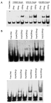
DNA binding properties of recombinant His6-tagged SASP4. A) Electromobility shift assays (EMSA) showing binding to biotin-labelled C. perfringens DNA by purified rSASP4 from F4969 (a CPE-positive nonfood-borne human GI disease strain that forms sensitive spores and produces an SASP4 variant with a Gly at residue 36), SM101 or 01E809 (two CPE-positive food poisoning isolates that form resistant spores and produce an SASP4 variant with Asp at residue 36). B) EMSA showing binding by purified SM101 rSASP4 or rSASP2 to (left) C. perfringens AT-rich biotin-labelled DNA or (right) biotin-labeled C. perfringens GC-rich DNA. Reproduced with permission from (42, 78).
Figure 5
Figure 5
Current model for the mechanism of action of CPE. CPE binds to claudin receptors to form small complexes. Those small complexes then oligomerize on the host cell surface to form an ∼450 kDa prepore known as CH-1. The prepore inserts into the membrane to form an active pore that alters host plasma membrane permeability for small molecules. As a result, calcium enters the cytoplasm and triggers either apoptosis (caused by low CPE doses, where there is a modest calcium influx) or oncosis (caused by high CPE doses, where there is a strong calcium influx). Reproduced with permission from (1).

References

    1. McClane BA, Robertson SL, Li J. Clostridium perfringens. In: Doyle MP, Buchanan RL, editors. Food Microbiology: Fundamentals and Frontiers. 4th. ASM press; Washington, D.C.: 2013. pp. 465–489.
    1. McClane BA, Uzal FA, Miyakawa MF, Lyerly D, Wilkins TD. The Enterotoxic Clostridia. In: Dworkin M, Falkow S, Rosenburg E, Schleifer H, Stackebrandt E, editors. The Prokaryotes. 3rd. Springer NY press; New York: 2006. pp. 688–752.
    1. Mallozzi M, Viswanathan VK, Vedantam G. Spore-forming Bacilli and Clostridia in human disease. Future Microbiol. 2010;5:1109–1123. - PubMed
    1. Amimoto K, Noro T, Oishi E, Shimizu M. A novel toxin homologous to large clostridial cytotoxins found in culture supernatant of Clostridium perfringens type C. Microbiology. 2007;153:1198–1206. - PubMed
    1. Stevens DL, Rood JI. Histotoxic Clostridia. In: Fischetti VA, Novick RP, Ferretti JJ, Portnoy DA, Rood JI, editors. Gram-positive pathogens. 2nd. ASM press; Washington, DC: 2006.

Publication types

MeSH terms

LinkOut - more resources