Review
. 2016 Jun;4(3):10.1128/microbiolspec.MCHD-0011-2015.
doi: 10.1128/microbiolspec.MCHD-0011-2015.

Osteoclasts-Key Players in Skeletal Health and Disease

Affiliations
Review

Osteoclasts-Key Players in Skeletal Health and Disease

Deborah Veis Novack et al. Microbiol Spectr. 2016 Jun.

Abstract

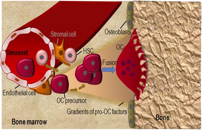
The differentiation of osteoclasts (OCs) from early myeloid progenitors is a tightly regulated process that is modulated by a variety of mediators present in the bone microenvironment. Once generated, the function of mature OCs depends on cytoskeletal features controlled by an αvβ3-containing complex at the bone-apposed membrane and the secretion of protons and acid-protease cathepsin K. OCs also have important interactions with other cells in the bone microenvironment, including osteoblasts and immune cells. Dysregulation of OC differentiation and/or function can cause bone pathology. In fact, many components of OC differentiation and activation have been targeted therapeutically with great success. However, questions remain about the identity and plasticity of OC precursors and the interplay between essential networks that control OC fate. In this review, we summarize the key principles of OC biology and highlight recently uncovered mechanisms regulating OC development and function in homeostatic and disease states.


Figures

Figure 1
Figure 1
A model of OC differentiation. OC differentiate from HSC. The hematopoietic niche comprises endothelial cells and perivascular stromal cells, which exhibit MSC features. It is still unclear whether OC precursors directly differentiate into OC or enter the bloodstream before re-entering the bone microenvironment to form OC. In any scenario, higher levels of chemoattractants toward bone surfaces, including bone extracellular matrix (ECM) proteins, lipid mediators (e.g., sphingosine-1-phosphate) and ECM degradation products create gradients that attract OC precursors to the hard tissue where they fuse and complete the differentiation process. Conversely, higher levels of peri-vascular chemorepellents (not drawn for simplicity) may also contribute to the migration of OC precursors toward the endosteum.
Figure 2
Figure 2
Key molecules involved in OC function. Loss-of-function of any of the depicted molecules causes osteopetrosis due to defective OC activity. OC adhere to bone matrix proteins via integrin αvβ3 and polarized such that the plasma membrane facing bone is convoluted (ruffled) and contains the proton pump (v-ATPase) and Cl channel 7 (ClC7), whereas the basolateral membrane is bears the HCO3/Cl antiporter. Cytoplasmic carbonic anhydrase type II (CAII) generates the protons to be secreted into the resorption lacuna beneath the cell. This lacuna becomes isolated from the rest of the extracellular space by the tight adhesion of αvβ3 to the bone surface at the sealing zone. The cytoplasmic domain of β3 recruits signaling proteins, which induce the association of actin with interacting partners (including talin, vinculin, kindlin, myosin IIA and paxillin), and formation of an actin ring that defines the periphery of the ruffled membrane. Concerted action of ClC7 and v-ATPase produces a high concentration of HCl that acidifies the resorption lacuna, leading to the dissolution of the inorganic components of the bone matrix. Acidified cytoplasmic vesicles containing lysosomal enzymes such as cathepsin K (Cat K) are also transported toward the bone-apposed plasma membrane, and ultimately, the sealed resorption lacuna, where they digest the exposed matrix proteins.

References

    1. Mosaad YM. Hematopoietic stem cells: an overview. Transfus Apheresis Sci. 2014;51:68–82. - PubMed
    1. Demulder A, Takahashi S, Singer FR, Hosking DJ, Roodman GD. Abnormalities in osteoclast precursors and marrow accessory cells in Paget’s disease. Endocrinology. 1993;133:1978–1982. - PubMed
    1. Demulder A, Suggs SV, Zsebo KM, Scarcez T, Roodman GD. Effects of stem cell factor on osteoclast-like cell formation in long-term human marrow cultures. J Bone Miner Res. 1992;7:1337–1344. - PubMed
    1. Bonar SL, Brydges SD, Mueller JL, McGeough MD, Pena C, Chen D, Grimston SK, Hickman-Brecks CL, Ravindran S, McAlinden A, Novack DV, Kastner DL, Civitelli R, Hoffman HM, Mbalaviele G. Constitutively activated NLRP3 inflammasome causes inflammation and abnormal skeletal development in mice. PLoS One. 2012;7:e35979. - PMC - PubMed
    1. Mediero A, Perez-Aso M, Cronstein BN. Activation of EPAC1/2 is essential for osteoclast formation by modulating NFκB nuclear translocation and actin cytoskeleton rearrangements. FASEB J. 2014;28:4901–4913. - PMC - PubMed

Publication types

LinkOut - more resources