

Adamantinoma of the distal femur diagnosed 5 years after initial surgery: a case report
- PMID: 27337984
- PMCID: PMC4918021
- DOI: 10.1186/s13256-016-0974-8
Adamantinoma of the distal femur diagnosed 5 years after initial surgery: a case report
Abstract
Background: Adamantinoma arising in the femur is extremely rare. We report a case of an adamantinoma occurring in the right medial femoral condyle that was diagnosed 5 years after the primary surgery.
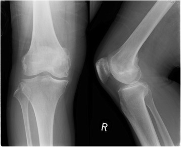
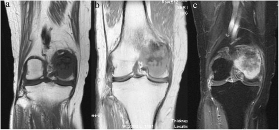
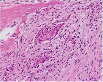
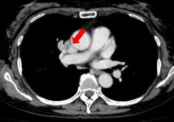
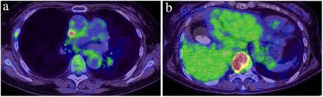
Case presentation: A 74-year-old Asian woman first complained of right knee pain without any cause. Radiographs demonstrated a 4×4.5 cm osteolytic lesion in her medial femoral condyle. Magnetic resonance imaging revealed a lesion which showed low signal on both T1 and T2-weighted image, and enhanced signal with gadolinium contrast administration. She underwent a wide resection of the lesion and was reconstructed with a tumor endoprosthesis. On histological examination, the tumor showed clusters of spindle-shaped and squamoid epithelial cells among the fibrous stroma. Adamantinoma was considered, however, the diagnosis was inconclusive due to the unusual localization and her age. Moreover, it was difficult to exclude metastatic carcinoma. Five years later, she was diagnosed with an abnormal shadow occupying the upper lobe of her right lung in a routine physical examination. She subsequently underwent a resection of the lung mass which histologically showed proliferation of spindle-shaped and squamoid epithelial cells. The histological similarity of the lung tumor and the femoral tumor led to the diagnosis of adamantinoma arising in her right medial femoral condyle with metastasis to the upper lobe of her right lung.
Conclusion: In this case report, we report the clinical, radiographic, and histological features of an adamantinoma arising in the distal femur with a review of the literature.
Keywords: Adamantinoma; Distal femur; Medial femoral condyle; Metastatic adamantinoma.
Figures


References
-
- Kitsoulis P, Charchati A, Paraskevas G, Marini A, Karatzias G. Adamantinoma. Acta Orthop Belg. 2007;73:425–31. - PubMed
-
- Hazelbag HM, Taminiau AH, Fleuren GJ, Hogendoorn PC. Adamantinoma of the long bones. A clinicopathological study of thirty-two patients with emphasis on histological subtype, precursor lesion, and biological behavior. J Bone Joint Surg Am. 1994;76:1482–99. - PubMed
-
- Chandrasekar CR, Mohammed R, Rafalla AA, Grimer RJ. Adamantinoma of the calcaneum – a case report. Foot(Edinb) 2009;19:58–61. - PubMed
Publication types
MeSH terms
LinkOut - more resources
Full Text Sources
Other Literature Sources
Medical
