. 2016 Sep;90(3):214-24.
doi: 10.1124/mol.116.104828. Epub 2016 Jun 23.

Dihydromunduletone Is a Small-Molecule Selective Adhesion G Protein-Coupled Receptor Antagonist

Affiliations

Dihydromunduletone Is a Small-Molecule Selective Adhesion G Protein-Coupled Receptor Antagonist

Hannah M Stoveken et al. Mol Pharmacol. 2016 Sep.

Abstract

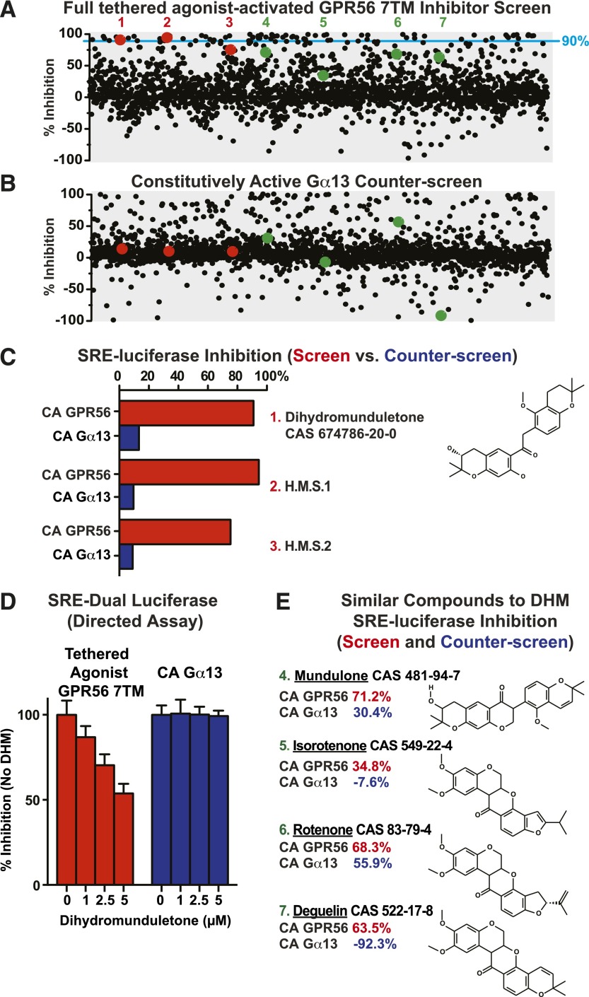
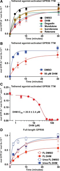
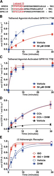
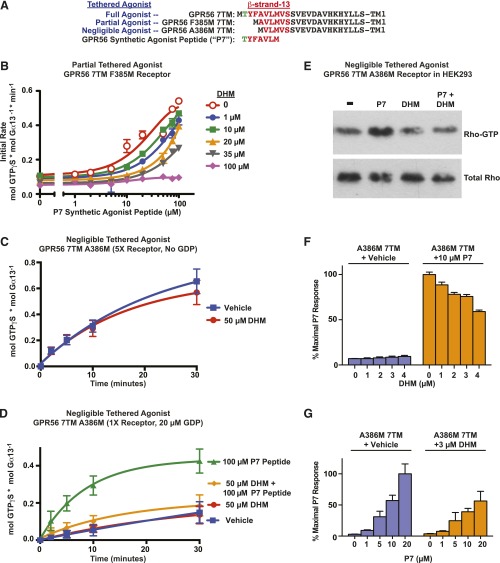
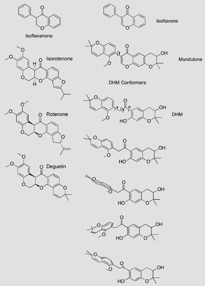
Adhesion G protein-coupled receptors (aGPCRs) have emerging roles in development and tissue maintenance and is the most prevalent GPCR subclass mutated in human cancers, but to date, no drugs have been developed to target them in any disease. aGPCR extracellular domains contain a conserved subdomain that mediates self-cleavage proximal to the start of the 7-transmembrane domain (7TM). The two receptor protomers, extracellular domain and amino terminal fragment (NTF), and the 7TM or C-terminal fragment remain noncovalently bound at the plasma membrane in a low-activity state. We recently demonstrated that NTF dissociation liberates the 7TM N-terminal stalk, which acts as a tethered-peptide agonist permitting receptor-dependent heterotrimeric G protein activation. In many cases, natural aGPCR ligands are extracellular matrix proteins that dissociate the NTF to reveal the tethered agonist. Given the perceived difficulty in modifying extracellular matrix proteins to create aGPCR probes, we developed a serum response element (SRE)-luciferase-based screening approach to identify GPR56/ADGRG1 small-molecule inhibitors. A 2000-compound library comprising known drugs and natural products was screened for GPR56-dependent SRE activation inhibitors that did not inhibit constitutively active Gα13-dependent SRE activation. Dihydromunduletone (DHM), a rotenoid derivative, was validated using cell-free aGPCR/heterotrimeric G protein guanosine 5'-3-O-(thio)triphosphate binding reconstitution assays. DHM inhibited GPR56 and GPR114/ADGRG5, which have similar tethered agonists, but not the aGPCR GPR110/ADGRF1, M3 muscarinic acetylcholine, or β2 adrenergic GPCRs. DHM inhibited tethered peptide agonist-stimulated and synthetic peptide agonist-stimulated GPR56 but did not inhibit basal activity, demonstrating that it antagonizes the peptide agonist. DHM is a novel aGPCR antagonist and potentially useful chemical probe that may be developed as a future aGPCR therapeutic.


Figures

Fig. 1.
Fig. 1.
A cell-based high-throughput screen and counterscreen to identify candidate small-molecule GPR56 inhibitors. HEK293T cells were cotransfected with the SRE-luciferase reporter plasmid and full tethered-agonist-activated GPR56 7TM pcDNA3.1 (A) or constitutively active GNA13Q226L pcDNA3.1 (B). Transfected cells were incubated overnight in 384-well format with ∼2000 individual compounds (5 µM each) from the Spectrum Collection. SRE reporter activity was measured in response to each compound and reported as percentage of inhibition over DMSO control for the screen and counterscreen. Top hits for individual compounds are represented as red, single dots, and compounds structurally similar to the top hits are represented as green dots. The 90% inhibition threshold is shown with a light-blue line. (C) Compounds 1 (DHM), 2 (H.M.S.1), and 3 (H.M.S.2) were identified as top hits from the GPR56 7TM inhibitor screen that did not appreciably inhibit the constitutively active (CA) Gα13-Q226L activity in the counterscreen. (D) Purchased dihydromunduletone was tested for concentration-dependent GPR56 7TM or Gα13-Q226L inhibition using a distinct, directed HEK293T cell SRE-luciferase assay. Error bars are the average ± the s.d. of three experimental replicates. (E) DHM-related flavonoid compounds present in the Spectrum Collection [4 (mundulone), 5 (isorotenone), 6 (rotenone), and 7 (deguelin)] all inhibited GPR56 7TM–activated SRE luciferase to a greater extent than Gα13-Q226L–activated SRE luciferase.
Fig. 2.
Fig. 2.
The rank potency of flavonoid derivative–induced HEK293 cell detachment correlates with electron transport chain complex I inhibition. (A) Adherent HEK293 cells were grown for 24 hours in the presence of the indicated concentrations of the five flavonoids (DHM, mundulone, rotenone, deguelin, isorotenone). Detached cells were removed and the relative levels of adhered cells remaining were measured by proxy assay of PicoGreen quantification of total DNA. (B) Isolated mitochondria were supplied with NADH and an electron acceptor (Q1), and the loss of NADH over 5 minutes at 340 nM was measured in the presence of the indicated amounts of each flavonoid. IC50 values were calculated from monoexponential association curves fitted using GraphPad Prism. R.F.U., relative fluorescent units. Error bars are the average ± the s.d. of three experimental replicates.
Fig. 3.
Fig. 3.
Secondary validation assays demonstrating that DHM and other rotenoids inhibit GPR56. (A–C) Prepared membranes containing the full tethered-agonist-activated GPR56 7TM receptor were reconstituted with purified G13 heterotrimer. The kinetics of G13 GTPγS binding were measured in the presence of 50 µM isoflavonoids (DHM, deguelin, mundulone, isorotenone, or rotenone) or DMSO vehicle control (A), or 50 µM DHM or DMSO control with 20 µM GDP present (B). Error bars are the average ± the s.d. of three experimental replicates. (C) The initial rates of full tethered-agonist-activated GPR56 7TM stimulation of G13 GTPγS binding were determined from four point (t = 0,1,2,3 min.) linear functions for each concentration of DHM. The IC50 of DHM inhibition was derived from the semilog plot of a one-phase monoexponential association function using GraphPad Prism. Error bars are the average of each linear line slope (rate) ± the s.d. of three technical replicates. (D) Membranes containing full-length (Karpus et al., 2013) GPR56 receptor were untreated or treated with ice-cold 7 M urea to mimic the proposed process of ligand-mediated receptor activation via N-terminal extracellular fragment dissociation. The kinetics of receptor-activated G13 GTPγS binding were measured in the presence of 50 µM DHM or DMSO control. Error bars were frequently smaller than the plotted symbols and are the average ± the s.d. of three experimental replicates. Full-length FL, full-length.
Fig. 4.
Fig. 4.
DHM exhibits specificity for group VIII adhesion GPCRs, but does not inhibit GPR110 or agonist-stimulated β2 adrenergic or M3 muscarinic acetylcholine class A GPCRs. (A) Alignment of the tethered agonist regions of group VI and group VIII adhesion GPCRs. Membranes containing the tethered-agonist-activated GPR114 7TM (B) or GPR110 7TM (C) receptors were reconstituted with purified Gs or Gq heterotrimers, respectively. The kinetics of heterotrimeric G protein GTPγS binding were measured in the presence of DHM (50 µM) or an equivalent volume of DMSO vehicle control. (D) M3 muscarinic acetylcholine receptor membranes were incubated with Gq and the indicated combinations of 50 µM carbachol (CCh), 50 µM DHM, or vehicle prior to measurement of Gq GTPγS binding kinetics. (E) β2 Adrenergic receptor membranes were incubated with Gs and the indicated combinations of 10 µM isoproterenol (ISO), 50 µM DHM, or vehicle prior to measurement of Gs GTPγS binding kinetics. Error bars are the average ± the s.d. of three experimental replicates.
Fig. 5.
Fig. 5.
DHM inhibits tethered- and synthetic-peptide agonist–stimulated GPR56, but does not inhibit basal receptor activity, a mode of action consistent with that of a neutral antagonist. (A) Alignment of stalk regions of intact tethered agonist and compromised tethered agonist GPR56 7TM domain receptors. The seven amino acid synthetic GPR56 agonist peptide is aligned underneath. (B) Partially compromised tethered agonist GPR56 F385M 7TM receptor membranes were reconstituted with G13, and P7 peptide concentration–dependent activation of G13 GTPγS binding was measured in response to increasing concentrations of DHM. Initial rates are plotted and were determined from four point (t = 0,1,2,3 min.) linear functions. Error bars are the average of each linear line slope (rate) ± the s.d. of three technical replicates. (C) The ability to measure near-basal activity of the GPR56 A386M 7TM receptor with a negligibly active tethered agonist was enhanced by use of five times more receptor membranes and excluding GDP from the reconstitution assay. GPR56 A386M 7TM basal activation of G13 GTPγS binding was measured in the presence of 50 µM DHM or the DMSO vehicle control. Error bars are the average ± the s.d. of three experimental replicates. (D) DHM inhibits P7 synthetic-peptide agonist stimulation of the compromised tethered agonist GPR56 A386M 7TM receptor. GPR56 A386M 7TM membranes were reconstituted with G13, and receptor-stimulated G13 GTPγS binding kinetics were measured in response to 100 µM P7 agonist peptide and/or 50 µM DHM in the presence of 20 µM GDP. Error bars are the average ± the s.d. of three experimental replicates. (E) P7 agonist-peptide (10 µM) stimulation of the GPR56 A386M 7TM receptor in intact HEK293 cells activated RhoA GTPase, an effect blocked by DHM (10 µM) (n = 3); shown is a representative immunoblot. (F) P7 agonist-peptide (10 µM) stimulation of the GPR56 A386M 7TM receptor in intact HEK293 cells activated SRE-luciferase. Increasing concentrations of DHM (1–4 µM) inhibited the P7 stimulus. Error bars are the average ± the s.d. of three technical replicates. (G) Increasing P7 agonist peptide (up to 20 µM) activated the SRE-luciferase reporter in a concentration-dependent manner, and 3 µM DHM inhibited P7 activation. Error bars are the average ± the s.d. of three experimental replicates.
Fig. 6.
Fig. 6.
Structural features of DHM and related compounds. The rotenoids (isorotenone, rotenone, and deguelin) are relatively rigid, planar compounds with isoflavanone-like cores. In contrast, mundulone, a true isoflavone, and DHM both lack the isoflavanone core, which allows for increased flexibility and the predicted ability to adopt multiple conformations. Mundulone may rotate around one central carbon-carbon bound, indicated by the arrow. The core of DHM is a 2-phenylacetophenone that would permit rotation about the three indicated carbon-carbon bonds, strongly suggesting that DHM is the most flexible compound of the five compounds studied. Representative conformers of DHM are shown to illustrate the predicted flexibility of the compound [conformational analysis was done with the Merck Molecular Force Field (MMFF) force field, iSpartan (www.wavefun.com); structures were drawn with ChemDraw Pro (Perkin Elmer, Waltham, MA)].

References

    1. Araç D, Boucard AA, Bolliger MF, Nguyen J, Soltis SM, Südhof TC, Brunger AT. (2012) A novel evolutionarily conserved domain of cell-adhesion GPCRs mediates autoproteolysis. EMBO J 31:1364–1378. - PMC - PubMed
    1. Arunlakshana O, Schild HO. (1959) Some quantitative uses of drug antagonists. Br Pharmacol Chemother 14:48–58. - PMC - PubMed
    1. Bindslev N(2008) The Schild against other theories, in Drug-Acceptor Interactions pp 283–298, Coaction Publishing, Stockholm.
    1. Bolliger MF, Martinelli DC, Südhof TC. (2011) The cell-adhesion G protein-coupled receptor BAI3 is a high-affinity receptor for C1q-like proteins. Proc Natl Acad Sci USA 108:2534–2539. - PMC - PubMed
    1. Boucard AA, Ko J, Südhof TC. (2012) High affinity neurexin binding to cell adhesion G-protein-coupled receptor CIRL1/latrophilin-1 produces an intercellular adhesion complex. J Biol Chem 287:9399–9413. - PMC - PubMed

Publication types

MeSH terms