Comparative Study
. 2016 Jun 22;13(6):619.
doi: 10.3390/ijerph13060619.

Comparison of Individual and Combined Effects of Four Endocrine Disruptors on Estrogen Receptor Beta Transcription in Cerebellar Cell Culture: The Modulatory Role of Estradiol and Triiodo-Thyronine

Affiliations
Comparative Study

Comparison of Individual and Combined Effects of Four Endocrine Disruptors on Estrogen Receptor Beta Transcription in Cerebellar Cell Culture: The Modulatory Role of Estradiol and Triiodo-Thyronine

Gergely Jocsak et al. Int J Environ Res Public Health. .

Abstract

Background: Humans and animals are continuously exposed to a number of environmental substances that act as endocrine disruptors (EDs). While a growing body of evidence is available to prove their adverse health effects, very little is known about the consequences of simultaneous exposure to a combination of such chemicals;

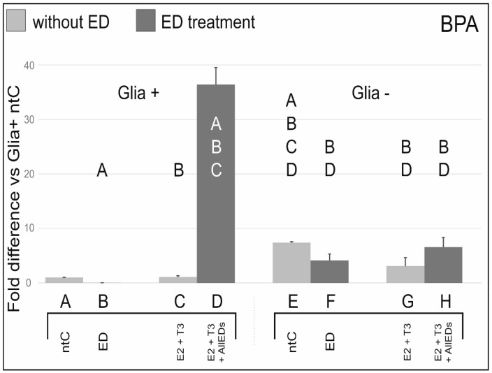
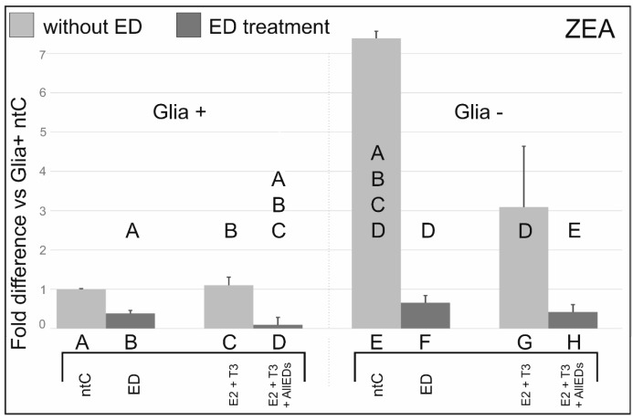
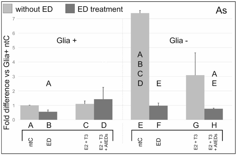
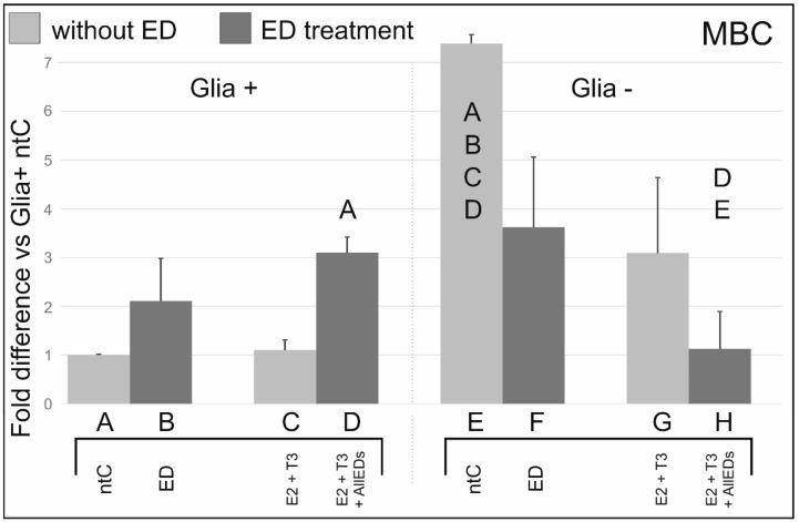
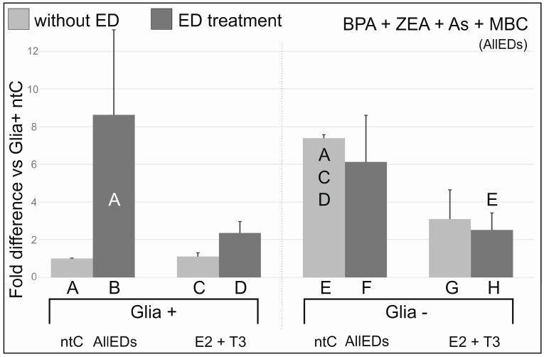
Methods: Here, we used an in vitro model to demonstrate how exposure to bisphenol A, zearalenone, arsenic, and 4-methylbenzylidene camphor, alone or in combination, affect estrogen receptor β (ERβ) mRNA expression in primary cerebellar cell cultures. Additionally, we also show the modulatory role of intrinsic biological factors, such as estradiol (E2), triiodo-thyronine (T3), and glial cells, as potential effect modulators;

Results: RESULTS show a wide diversity in ED effects on ERβ mRNA expression, and that the magnitude of these ED effects highly depends on the presence or absence of E2, T3, and glial cells;

Conclusion: The observed potency of the EDs to influence ERβ mRNA expression, and the modulatory role of E2, T3, and the glia suggests that environmental ED effects may be masked as long as the hormonal milieu is physiological, but may tend to turn additive or superadditive in case of hormone deficiency.

Keywords: arsenic; bisphenol A; camphor; combined treatment; estrogen receptor beta; transcription; zearalenone.


Figures

Figure 1
Figure 1
Effects of bisphenol A (BPA) with, and without, 17beta-estradiol (E2) and triiodo-thyronine (T3), on ERβ mRNA expression. BPA treatment decreased ERβ mRNA expression compared to the non-treated control. In glia-reduced cultures (Glia−), a more than two orders of magnitude higher (×180) ERβ mRNA expression was found in BPA treated cultures (column B vs. F). In glia containing samples (Glia+), there was about three magnitudes higher ERβ mRNA expression after E2 + T3 + BPA (column D) treatment than after BPA treatment alone (column B). Letters in and above the columns indicate significant differences (p ≤ 0.05) from columns below.
Figure 2
Figure 2
Effects of zearalenone (Zea) with, and without, 17beta-estradiol (E2) and triiodo-thyronine (T3), on ERβ mRNA expression. ERβ mRNA expression was substantially reduced by Zea treatment compared to the respective ntCs and Zea-void hormone treated cultures. Letters in and above the columns indicate significant differences (p ≤ 0.05) from columns below.
Figure 3
Figure 3
Effects of arsenic (As) with, and without, 17beta-estradiol (E2) and triiodo-thyronine (T3), on ERβ mRNA expression. ERβ mRNA expression levels after As and As + E2 + T3 treatments showed a pattern comparable in trends with that found in BPA studies in Glia+ and to those seen in Zea studies in Glia−. Letters in and above the columns indicate significant differences (p ≤ 0.05) from columns below.
Figure 4
Figure 4
Effects of methylbenzylidene camphor (MBC) with, and without, 17beta-estradiol (E2) and triiodo-thyronine (T3), on ERβ mRNA expression. MBC tended to increase ERβ mRNA expression in Glia+ cultures. In Glia−, MBC decreased ERβ mRNA compared to its respective ntC, and E2 + T3 treated counterparts, although these differences did not reach significance. Letters in and above the columns indicate significant differences (p ≤ 0.05) from columns below.
Figure 5
Figure 5
Effects of co-treatments with bisphenol A (BPA), zearalenone (Zea), arsenic (As), and methylbenzylidene camphor (MBC) with, and without 17beta-estradiol (E2) and triiodo-thyronine (T3) on ERβ mRNA expression. There was a robust and significant increase in ERβ mRNA expression in AllEDs treatment group of glia containing (Glia+) cultures, when compared to the ntC. However, high variance in differences were also found. In contrast, no increase was found in glia-reduced (Glia−) samples. ERβ mRNA expression values in E2 + T3 + All EDs groups were nearly identical in Glia+ vs. Glia− cultures. Compared to their respective non-treated controls, transcript levels in E2 + T3 + AllEDs cultures doubled in Glia+, while decreased to the one third in Glia−. Letters above the columns indicate significant differences (p ≤ 0.05) from columns below.

References

    1. Zsarnovszky A., Földvári E.G., Rónai Z., Bartha T., Frenyó L.V. Oestrogens in the mammalian brain: From conception to adulthood—A review. Acta Vet. Hung. 2007;55:333–347. doi: 10.1556/AVet.55.2007.3.8. - DOI - PubMed
    1. Jakab R.L., Wong J.K., Belcher S.M. Estrogen receptor beta immunoreactivity in differentiating cells of the developing rat cerebellum. J. Comp. Neurol. 2001;430:396–409. doi: 10.1002/1096-9861(20010212)430:3<396::AID-CNE1039>3.0.CO;2-0. - DOI - PubMed
    1. Ikeda Y. Expression of the two estrogen receptor (ER) subtypes, ERalpha and ERbeta, during postnatal development of the rat cerebellum. Cerebellum. 2008;7:501–502. - PubMed
    1. Fan X., Xu H., Warner M., Gustafsson J.-A. ERbeta in CNS: New roles in development and function. Prog. Brain Res. 2010;181:233–250. - PubMed
    1. Belcher S.M. Regulated expression of estrogen receptor alpha and beta mRNA in granule cells during development of the rat cerebellum. Brain Res. Dev. Brain Res. 1999;115:57–69. doi: 10.1016/S0165-3806(99)00050-4. - DOI - PubMed

Publication types

LinkOut - more resources