Review
. 2016 Jun 7;8(6):160.
doi: 10.3390/v8060160.

Endoplasmic Reticulum: The Favorite Intracellular Niche for Viral Replication and Assembly

Affiliations
Review

Endoplasmic Reticulum: The Favorite Intracellular Niche for Viral Replication and Assembly

Inés Romero-Brey et al. Viruses. .

Abstract

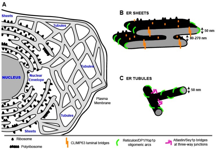
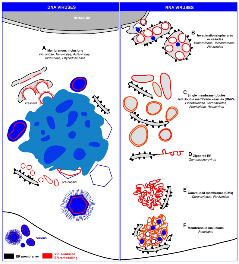
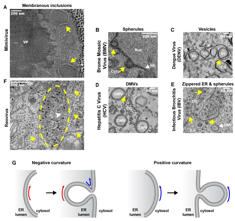
The endoplasmic reticulum (ER) is the largest intracellular organelle. It forms a complex network of continuous sheets and tubules, extending from the nuclear envelope (NE) to the plasma membrane. This network is frequently perturbed by positive-strand RNA viruses utilizing the ER to create membranous replication factories (RFs), where amplification of their genomes occurs. In addition, many enveloped viruses assemble progeny virions in association with ER membranes, and viruses replicating in the nucleus need to overcome the NE barrier, requiring transient changes of the NE morphology. This review first summarizes some key aspects of ER morphology and then focuses on the exploitation of the ER by viruses for the sake of promoting the different steps of their replication cycles.

Keywords: cell membranes; electron microscopy; intracellular organelles; membrane rearrangements; nuclear envelope; peripheral endoplasmic reticulum (ER); vesicles; viral replication; virion assembly; virus-host interactions.


Figures

Figure 1
Figure 1
Schematic representation of the endoplasmic reticulum (ER) network organization. (A) The ER network comprises the nuclear envelope (NE) and the peripheral ER. The peripheral ER is composed of stacked sheets, studded with ribosomes, and tubules extending the reticular network to the plasma membrane; (B) Schematic of an ER sheet, depicting its multidimensional properties and the ER-shaping proteins that are responsible for the characteristic, flattened morphology. CLIMP63 is a coiled-coil protein forming oligomers bridging across the luminal space of the sheets that are also studded with ribosomes. High membrane curvature at the edges of the sheets is stabilized by reticulon/DP1/Yop1p proteins, forming wedges inside the bilayer and arc-like scaffolds around the edge. Sheets are also connected to neighboring sheets by membranous twists (“Parking garage model”, [7]); (C) Schematic of two ER tubules. Reticulon/DP1/Yop1p oligomers might not only determine the size of the sheet edges, but also the diameter of the tubules. These are interconnected through proteins such as atlastins/Sey1p residing at three-way junctions. The models in (B) and (C) are adapted from [8,9]. Dimensions of ER sheets and tubules refer to those found in mammalian cells.
Figure 2
Figure 2
Schematic representation of ER modifications induced by DNA and RNA viruses (left and right panel, respectively). (A) Some DNA viruses (e.g., Poxviridae, Mimiviridae, Asfarviridae, Iridoviridae and Phycodnaviridae) replicate their genomes in the cytoplasm in close association with ER-derived membranes that are part of the replication and assembly factory. The single internal membrane bilayer of these viruses is generated through an unusual mechanism, comprising the generation of membrane sheets by rupture of ER cisternae in close proximity to the viral factories. There the single bilayers interact with capsid proteins to generate crescents and eventually pre-capsids. These factories have an ordered organization, with a preference to accumulate the viral genomes in the factory center to facilitate continuous generation of virus progeny. The only exception known so far is PBCV-1 (not shown) that accumulates the viral genome at the periphery, while the single-membrane bilayers localize to the center of the viral factory; (B) Some RNA viruses replicate their genomes in spherules (families Bromoviridae and Tombusviridae) or vesicles (family Flaviviridae), that are formed as invaginations towards the ER lumen; (C) Replication factories originating from “exvaginations” from the ER, giving rise to large single-membrane tubules or double-membrane vesicles (DMVs) (e.g., several members of the families Picornaviridae, Coronaviridae, Arteriviriridae, Hepacivirus); (D) Infectious bronchitis virus (IBV, genus Gammacoronavirus) induces mainly zippered ER membranes comprising tethered spherules. IBV also induces DMVs, albeit to low numbers; (E) ER-derived convoluted membranes (CMs) are also found in cells infected with members of the Coronaviridae and Flaviviridae. Since these structures are devoid of ribosomes, they are most likely modified smooth ER membranes; (F) In reovirus-infected cells, filled and empty virions (Reoviridae) are found embedded in a membranous inclusion of cellular origin (also called “viroplasm”) and closely surrounded by rough ER cisternae. Both ER elements and virus particles are in contact with the cytosolic face of the plasma membrane. Note that for reasons of simplicity, non-ER intracellular organelles are not shown in this schematic.
Figure 3
Figure 3
Representative electron micrographs of specialized virus-induced ER modifications. (A) Cytoplasmic viral factory (VF) of Acanthamoeba polyphaga mimivirus, 8 hpi, showing icosahedral capsids (yellow arrows) at the factory periphery; (B) The replication factor 1A of brome mosaic virus (BMV) localizes to outer perinuclear ER membranes in yeast cells, where it induces the formation of luminal spherules (yellow arrows) (60–80 nm) between the nucleoplasm (Nuc) and the cytoplasm (Cyto); (C) Dengue virus (DENV) induces the formation of invaginations of ER membranes, generating arrays of vesicles (Ve) that are known as vesicle packets (VPs). These vesicles remain connected to the cytosol through 10 nm diameter pores (yellow arrow). Shown are Huh7 cells 24 hpi; (D) DMVs are abundantly found in Huh 7.5 cells infected with hepatitis C virus (HCV), 16 hpi. Some of them remain connected to their source organelle, the ER (yellow arrow); (E) Zippered ER membranes are found in CK cells infected with the gammacoronavirus infectious bronchitis virus (IBV), 16 hpi. The zippered membranes fold to form individual spherules with a channel connecting the cytosol (yellow arrows). DMVs are also found, but to a lesser extent; (F) Membranous inclusions (yellow dashed line) found in HeLa cells infected with the reovirus strain T3-T1M1, 12 hpi. Rough ER cisternae (yellow arrows) are found surrounding the inclusions that contain many virions and coated microtubuli inside (white arrowhead); (G) Schematic representation of membrane curvature types. Negative membrane curvature results in the formation of invaginations towards the ER lumen, generating spherules or luminal vesicles. Positive membrane curvature generates exvaginations of ER membranes towards the cytosol, giving rise to cytoplasmic vesicles, tubules or DMVs. Electron micrographs are reproduced with permission from [76] (A), [150] (B), [146] (C,D) [107] (E) and [80] (F).

References

    1. Schuldiner M., Schwappach B. From rags to riches—The history of the endoplasmic reticulum. Biochim. Biophys. Acta. 2013;1833:2389–2391. doi: 10.1016/j.bbamcr.2013.03.005. - DOI - PubMed
    1. Veratti E. Investigations on the fine structure of striated muscle fiber read before the reale istituto lombardo, 13 March 1902. J. Biophys. Biochem. Cytol. 1961;10:1–59. doi: 10.1083/jcb.10.4.1. - DOI - PMC - PubMed
    1. Porter K.R. Observations on a submicroscopic basophilic component of cytoplasm. J. Exp. Med. 1953;97:727–750. doi: 10.1084/jem.97.5.727. - DOI - PMC - PubMed
    1. Palade G.E., Porter K.R. Studies on the endoplasmic reticulum. I. Its identification in cells in situ. J. Exp. Med. 1954;100:641–656. doi: 10.1084/jem.100.6.641. - DOI - PMC - PubMed
    1. Porter K.R., Palade G.E. Studies on the endoplasmic reticulum. III. Its form and distribution in striated muscle cells. J. Biophys. Biochem. Cytol. 1957;3:269–300. doi: 10.1083/jcb.3.2.269. - DOI - PMC - PubMed

Publication types

LinkOut - more resources