Review
. 2016 Jun 7;6(2):28.
doi: 10.3390/biom6020028.

NMR Meets Tau: Insights into Its Function and Pathology

Affiliations
Review

NMR Meets Tau: Insights into Its Function and Pathology

Guy Lippens et al. Biomolecules. .

Abstract

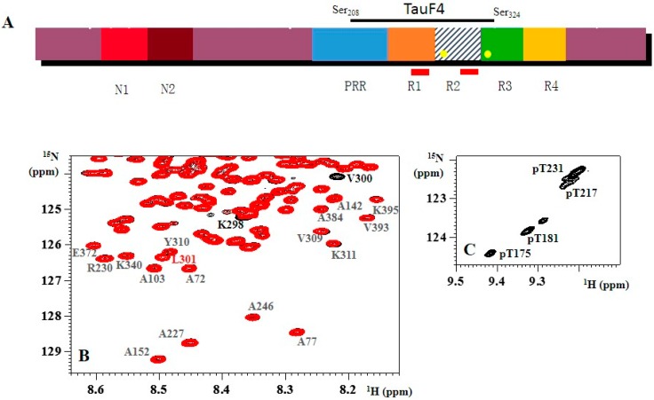
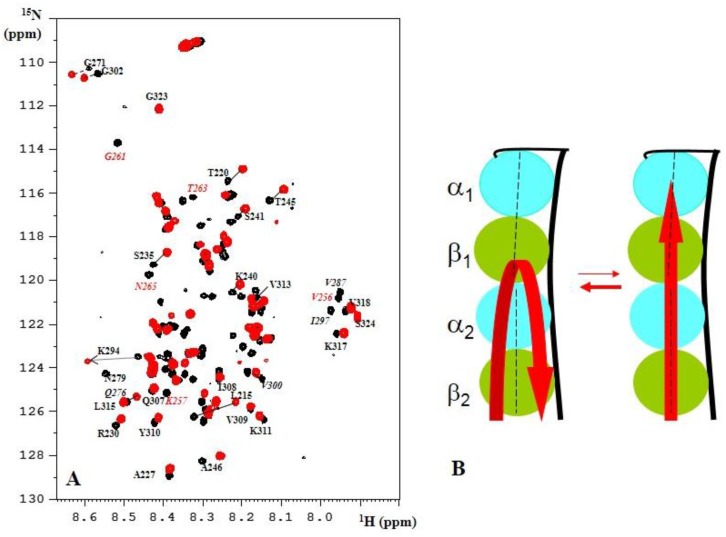
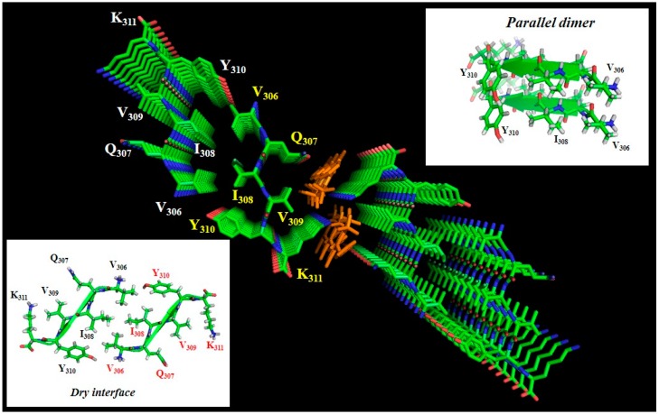
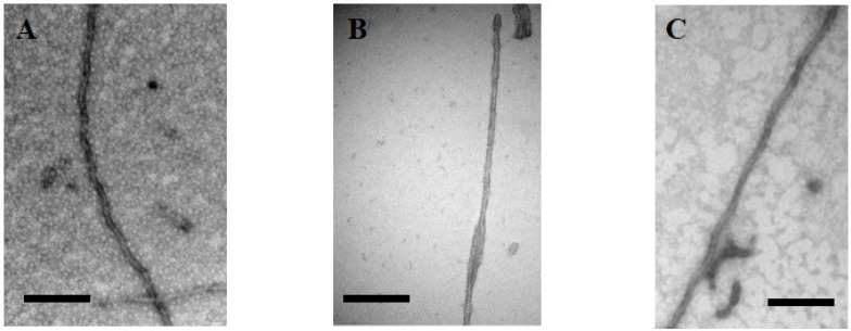
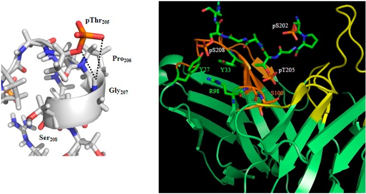
In this review, we focus on what we have learned from Nuclear Magnetic Resonance (NMR) studies on the neuronal microtubule-associated protein Tau. We consider both the mechanistic details of Tau: the tubulin relationship and its aggregation process. Phosphorylation of Tau is intimately linked to both aspects. NMR spectroscopy has depicted accurate phosphorylation patterns by different kinases, and its non-destructive character has allowed functional assays with the same samples. Finally, we will discuss other post-translational modifications of Tau and its interaction with other cellular factors in relationship to its (dys)function.

Keywords: NMR spectroscopy; Tau; aggregation; intrinsically disordered protein; phosphorylation; protein/protein interactions; tubulin.


Figures

Figure 1
Figure 1
(A) Schematic view of the primary sequence of Tau441, the longest isoform of Tau. Different isoforms are characterized by the insertion of 0, one or two N-terminal inserts (N1 and N2), and three (R1-R3-R4) or four (R1-4) repeats (leading to the 3R or 4R forms). The repeats are preceded by a proline-rich region (PRR). The two hexapeptides PHF6 and PHF6* are indicated as red rectangles in R1 and R2, whereas Tau’s two cysteine residues (Cys291 and Cys322) are indicated as yellow circles in R2 and R3. The fragment TauF4 spans part of the PRR, the first two repeats and a small part of R3; (B) The 1H, 15N HSQC spectra of 15N-labeled wild-type (black) and 15N,13C-labeled P301L (red) Tau441 show that only a couple of residues directly adjacent to the mutation show chemical shift differences. Residues Val309-Tyr310-Lys311 of the PHF6 peptide are identical in chemical shift and intensity in both proteins; (C) Zoom of the 1H, 15N spectrum of CDK2 phosphorylated Tau around the resonance of pThr231, showing several peaks for the same phosphorylated residue.
Figure 2
Figure 2
(A) The 1H, 15N TROSY spectrum of TauF4 in its complex with T2R. Residues in the R1 repeat whose resonances are absent in the TauF4-T2R spectrum are labeled in red; (B) Model of the swing movement of TauF4 on the curved T2R surface [76].
Figure 3
Figure 3
Crystal structure of the PHF6 peptide in complex with orange G (PDB code 3OVL), seen along the fiber axis from the top. Inserts show the antiparallel dry interface between opposing peptides, or the parallel stacking between adjacent peptides in the same plane. Orange G has two sulfate groups separated by 5 Å, and thereby compensates the lysine side-chain positive charges of two adjacent peptides [120].
Figure 4
Figure 4
Electron microscopy of (A) heparin-induced synthetic fibers of Tau441 show the morphology of paired helical filaments, whereas (B) those of a TauF4 fragment devoid of cysteine residues show a morphology of two flat ribbons twisted around one another [126]. This morphology is found in the ex vivo 3R fibers characterizing Pick’s disease [127]; (C) TauP301L after phosphorylation by Erk2 forms fibers without heparin. Scale bar = 100 nm.
Figure 5
Figure 5
Structure of the AT8 motif in solution [56] or in complex with the AT8 Fab fragment ([142]; PDB code 5E2W). The helical turn stabilized by the interaction between the Gly207 HN and the phosphate group of pThr205 (left) is not maintained in its complex with the AT8 antibody (right). The phosphate group of pSer208 makes additional interactions with residues of the antibody. The AT8 Fab is in green, with the variable loops of the light and heavy chain in yellow, respectively, orange.

References

    1. Weingarten M.D., Lockwood A.H., Hwo S.Y., Kirschner M.W. A protein factor essential for microtubule assembly. Proc. Natl. Acad. Sci. USA. 1975;72:1858–1862. doi: 10.1073/pnas.72.5.1858. - DOI - PMC - PubMed
    1. Brion J.P., Flament-Durand J., Dustin P. Alzheimer’s disease and tau proteins. Lancet. 1986;2 doi: 10.1016/S0140-6736(86)90495-2. - DOI - PubMed
    1. Grundke-Iqbal I., Iqbal K., Quinlan M., Tung Y.C., Zaidi M.S., Wisniewski H.M. Microtubule-associated protein tau. A component of Alzheimer paired helical filaments. J. Biol. Chem. 1986;261:6084–6089. - PubMed
    1. Wood J.G., Mirra S.S., Pollock N.J., Binder L.I. Neurofibrillary tangles of Alzheimer disease share antigenic determinants with the axonal microtubule-associated protein tau (tau) Proc. Natl. Acad. Sci. USA. 1986;83:4040–4043. doi: 10.1073/pnas.83.11.4040. - DOI - PMC - PubMed
    1. Kosik K.S., Joachim C.L., Selkoe D.J. Microtubule-associated protein tau (tau) is a major antigenic component of paired helical filaments in Alzheimer disease. Proc. Natl. Acad. Sci. USA. 1986;83:4044–4048. doi: 10.1073/pnas.83.11.4044. - DOI - PMC - PubMed

Publication types

MeSH terms

LinkOut - more resources