. 2016 Jun 23;9(1):67.
doi: 10.1186/s13041-016-0248-9.

Presynaptic muscarinic acetylcholine autoreceptors (M1, M2 and M4 subtypes), adenosine receptors (A1 and A2A) and tropomyosin-related kinase B receptor (TrkB) modulate the developmental synapse elimination process at the neuromuscular junction

Affiliations

Presynaptic muscarinic acetylcholine autoreceptors (M1, M2 and M4 subtypes), adenosine receptors (A1 and A2A) and tropomyosin-related kinase B receptor (TrkB) modulate the developmental synapse elimination process at the neuromuscular junction

Laura Nadal et al. Mol Brain. .

Abstract

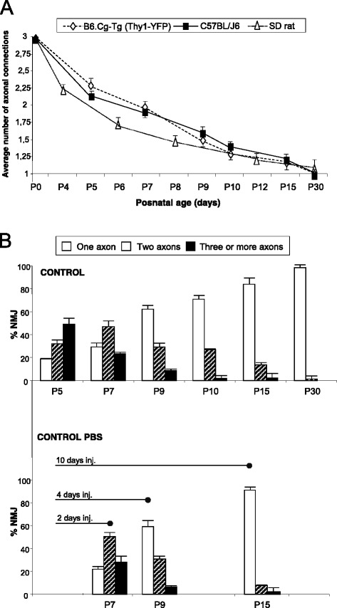
Background: The development of the nervous system involves an initially exuberant production of neurons that make an excessive number of synaptic contacts. The initial overproduction of synapses promotes connectivity. Hebbian competition between axons with different activities (the least active are punished) leads to the loss of roughly half of the overproduced elements and this refines connectivity and increases specificity. The neuromuscular junction is innervated by a single axon at the end of the synapse elimination process and, because of its relative simplicity, has long been used as a model for studying the general principles of synapse development. The involvement of the presynaptic muscarinic ACh autoreceptors may allow for the direct competitive interaction between nerve endings through differential activity-dependent acetylcholine release in the synaptic cleft. Then, the most active ending may directly punish the less active ones. Our previous results indicate the existence in the weakest axons on the polyinnervated neonatal NMJ of an ACh release inhibition mechanism based on mAChR coupled to protein kinase C and voltage-dependent calcium channels. We suggest that this mechanism plays a role in the elimination of redundant neonatal synapses.

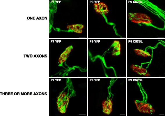
Results: Here we used confocal microscopy and quantitative morphological analysis to count the number of brightly fluorescent axons per endplate in P7, P9 and P15 transgenic B6.Cg-Tg (Thy1-YFP)16 Jrs/J mice. We investigate the involvement of individual mAChR M1-, M2- and M4-subtypes in the control of axonal elimination after the Levator auris longus muscle had been exposed to agonist and antagonist in vivo. We also analysed the role of adenosine receptor subtypes (A1 and A2A) and the tropomyosin-related kinase B receptor. The data show that postnatal axonal elimination is a regulated multireceptor mechanism that guaranteed the monoinnervation of the neuromuscular synapses.

Conclusion: The three receptor sets considered (mAChR, AR and TrkB receptors) intervene in modulating the conditions of the competition between nerve endings, possibly helping to determine the winner or the lossers but, thereafter, the final elimination would occur with some autonomy and independently of postsynaptic maturation.

Keywords: Cholinergic synapses; Motor end-plate; Motor nerve terminal; Neuromuscular junction.


Figures

Fig. 1
Fig. 1
The picture shows some representative confocal immunofluorescence images of the singly- and polyinnervated NMJ from YFP and C57BL/J6 mice. Scale bar: 10 μm
Fig. 2
Fig. 2
Postnatal evolution of polyneuronal innervation. In a, comparison of the results of axon counts in fluorencence immunohistochemistry LAL preparations of YFP and C57BL/J6 mice. The histogram in b shows the percentage of singly-, dually- and triply- (or more) innervated synapses in YFP animals on the postnatal days studied without any experimental manipulation (control non-PBS, without subcutaneous injection), and also at P7, P9 and P15 after two (days 5–6), four (days 5–8) and ten (days 5–14) daily subcutaneous PBS applications respectively (control PBS). No differences are observed between PBS and non-PBS preparations (Fisher’s test: p > 0,05)
Fig. 3
Fig. 3
Changes in polyneuronal innervation of the NMJ after stimulation and inhibition of the mAChR. The figure shows the percentage of singly-, dually- and triply- (or more) innervated NMJs in the untreated YFP control mice (exposed to PBS applications) and after 2 (P7 in a), 4 (P9, in b) and in some cases 10 (P15, in c) applications (one application every day after P5) of the mAChR agonist (ago.) oxotremorine (OXO) and such antagonists (ant.) as atropine (AT), pirenzepine (PIR), methoctramine (MET) and muscarinic toxin 3 (MT3). Fisher’s test: * p < 0,05, ** p < 0,01, *** p < 0,005
Fig. 4
Fig. 4
Involvement of adenosine receptors (AR) and TrkB receptors in axonal elimination. The figure shows the percentage of the singly-, dually- and triply- (or more) innervated NMJs in the YFP control mice exposed to PBS, and after 2 (P7 in a), 4 (P9, in b) and in some cases 10 (P15, in c) applications (one application every day after P5) of the BDNF-TrkB blocking pathway agent TrkB-Fc to sequester endogenous BDNF/NT-4 neurotrophins and the AR pan-inhibitor 8SPT and the AR agonist (ago.) ADO. We also studied axonal elimination after selectively blocking A1R with the antagonist (ant.) DPCPX and inhibiting A2AR with SCH-58261. The control for these selective inhibitors was PBS + DMSO (not shown in the figure) which shows no differences from PBS used as a control by itself. Fisher’s test * p < 0,05, ** p < 0,01, *** p < 0,005
Fig. 5
Fig. 5
Postnatal morphological maturation of the postsynaptic apparatus. The axonal elimination process is accompanied by changes in the morphology of the nAChR clusters in the postsynaptic membrane. a, the following maturation stages (MS1–MS4) were defined. MS1: Uniform nAChR oval plaque with an indistinct boundary seen in the majority of NMJs at birth. A uniformly distributed porosity can be observed within this plaque. MS2: nAChR elongated oval plaque with a few hints of inhomogeneities in receptor density. The nAChRs are denser on a few narrow ridges that occur within the plaque. MS3: An oval nAChR plaque with one or more fluorescence-free “holes.” These holes are not innervated. MS4: The oval nAChR areas have been transformed into a more mature branched pattern with a moderately convoluted external border and high and low receptor density areas. The edge of the holes usually has a high density of receptors. Scale bar: 10 μm. b, shows the percentages of the MS1-MS4 nAChR clusters plotted in the interval P5-P15 days
Fig. 6
Fig. 6
Maturation of postsynaptic nAChR clusters after stimulation and inhibition of mAChRs. Percentage of MS1-MS4 clusters in the NMJ of untreated YFP control mice (exposed to PBS), and after 2 (P7, in a), 4 (P9, in b) and in some cases 10 (P15 in c) applications of the muscarinic substances considered: OXO, AT, PIR, MET and MT3. Fisher’s test * p < 0,05, ** p < 0,01, *** p < 0,005
Fig. 7
Fig. 7
Pre- and postsynaptic maturation in the MS3 clusters after stimulation and inhibition of the mAChRs. For each day considered (P7 in a, P9 in b and P15 in c) the figure shows the percentage of MS3 clusters (the oval nAChR plaques with fluorescence-free holes that mature at a faster rate) with one, two and three or more axons as an indication of the appropiate correspondence of the pre- and postsynaptic maturation. Fisher’s test * p < 0,05, ** p < 0,01, *** p < 0,005
Fig. 8
Fig. 8
Involvement of the AR and TrkB receptors in the morphological maturation of the postsynaptic apparatus. The figure shows the percentage of the MS1-MS4 clusters in the NMJ of the untreated YFP control mice (exposed to PBS), and after 2 (P7 in a), 4 (P9, in b) and in some cases 10 (P15, in c) applications (one application every day after P5) of the TrkB blocking chimera TrkB-Fc, the AR pan-inhibitor 8SPT and the AR agonist ADO. We also studied axonal elimination after selectively blocking A1R with DPCPX and inhibiting A2AR with SCH-58261. Fisher’s test: * p < 0,05, ** p < 0,01, *** p < 0,005
Fig. 9
Fig. 9
Pre- and postsynaptic maturation in the MS3 clusters. AR and TrkB pathways modification. For each day considered (P7 in a, P9 in b and P15 in c), the figure shows the percentage of MS3 clusters with one, two and three or more axons as an indication of the appropiate correspondence of the pre- and postsynaptic maturation. Fisher’s test: * p < 0,05, ** p < 0,01, *** p < 0,005
Fig. 10
Fig. 10
Influence of the mAChR, AR and TrkB receptors on postnatal axonal elimination and synaptic maturation

Similar articles

Cited by

References

    1. Jansen JK, Fladby T. The perinatal reorganization of the innervation of skeletal muscle in mammals. Progress in Neurobiology. 1990;34:39–90. doi: 10.1016/0301-0082(90)90025-C. - DOI - PubMed
    1. Sanes JR, Lichtman JW. Development of the vertebrate neuromuscular junction. Annual Review of Neuroscience. 1999;22:389–442. doi: 10.1146/annurev.neuro.22.1.389. - DOI - PubMed
    1. Purves D, Lichtman JW. Elimination of synapses in the developing nervous system. Science. 1980;210:153–7. doi: 10.1126/science.7414326. - DOI - PubMed
    1. Chen C, Regehr WG. Developmental remodeling of the retinogeniculate synapse. Neuron. 2000;28:955–66. doi: 10.1016/S0896-6273(00)00166-5. - DOI - PubMed
    1. Ribchester RR, Barry JA. Spatial versus consumptive competition at polyneuronally innervated neuromuscular junctions. Experimental Physiology. 1994;79:465–94. doi: 10.1113/expphysiol.1994.sp003781. - DOI - PubMed

MeSH terms