

Slowly eroding lesions in multiple sclerosis
- PMID: 27339071
- PMCID: PMC5182188
- DOI: 10.1177/1352458516655403
Slowly eroding lesions in multiple sclerosis
Abstract
Background: At autopsy, 20%-40% of chronic multiple sclerosis (MS) lesions are labeled "slowly expanding" and feature myelin phagocytosis at the lesion edge. As pathological lesion classification relies on a single, terminal time point, the rate of lesion expansion cannot be directly measured.
Objective: To study long-term volume changes in individual MS lesions.
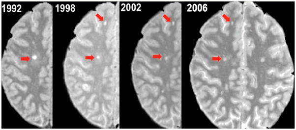
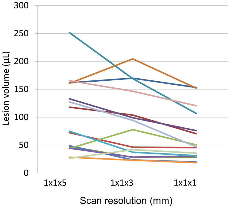
Methods: Volumes of individual lesions on proton density magnetic resonance imaging (MRI) acquired between 1992 and 2015 were measured in 22 individuals (one lesion per person). After correction for acquisition protocol, a mixed model evaluated lesion volume changes.
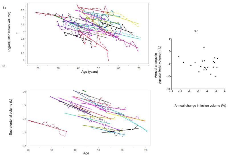
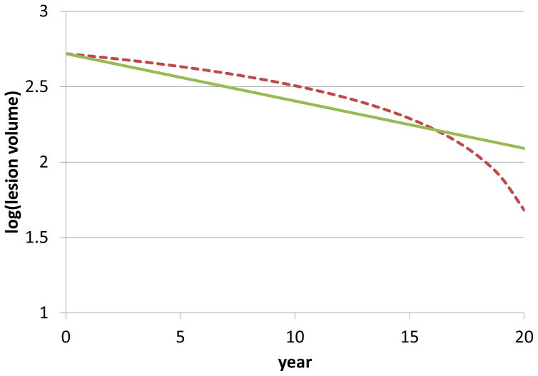
Results: The mean (standard deviation) lesion volume at baseline was 142 (82) mL, falling to 74 (51) mL after 16 (3) years. All lesions shrank over time. Change in lesion volume did not correlate with change in supratentorial brain volume ( p = 0.33). In simulations, the results could be explained by a process of slow radial expansion superimposed on substantially more rapid resorption of damaged tissue.
Conclusion: We noted sustained radiological contraction of MS lesions, a surprising result given that fresh myelin breakdown products within chronic active lesions are observed relatively frequently at autopsy. Therefore, the primary pathological process in chronic lesions, even those described as "slowly expanding," is likely to be tissue loss.
Keywords: Slowly expanding lesion; magnetic resonance imaging; multiple sclerosis; proton density.
Figures
References
-
- Dawson J. The histology of disseminated sclerosis. Trans R Soc Edinburgh. 1916 Jan 1;50:517–740.
-
- Lassmann H. The Pathologic Substrate of Magnetic Resonance Alterations in Multiple Sclerosis. Neuroimaging Clinics of North America. 2008 Nov;18(4):563–76. - PubMed
-
- Prineas JW, Kwon EE, Cho ES, Sharer LR, Barnett MH, Oleszak EL, et al. Immunopathology of secondary-progressive multiple sclerosis. Ann Neurol. 2001 Nov;50(5):646–57. - PubMed
-
- Yousry TA, Seelos K, Mayer M, Brüning R, Uttner I, Dichgans M, et al. Characteristic MR Lesion Pattern and Correlation of T1 and T2 Lesion Volume with Neurologic and Neuropsychological Findings in Cerebral Autosomal Dominant Arteriopathy with Subcortical Infarcts and Leukoencephalopathy (CADASIL) American Journal of Neuroradiology. 1999;20:91–100. - PubMed
MeSH terms
Grants and funding
LinkOut - more resources
Full Text Sources
Other Literature Sources
Medical
Research Materials
