. 2016 Jul 12;113(28):7858-63.
doi: 10.1073/pnas.1607769113. Epub 2016 Jun 23.

GsdmD p30 elicited by caspase-11 during pyroptosis forms pores in membranes

Affiliations

GsdmD p30 elicited by caspase-11 during pyroptosis forms pores in membranes

Robin A Aglietti et al. Proc Natl Acad Sci U S A. .

Abstract

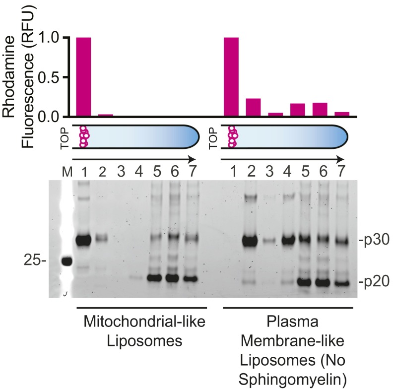
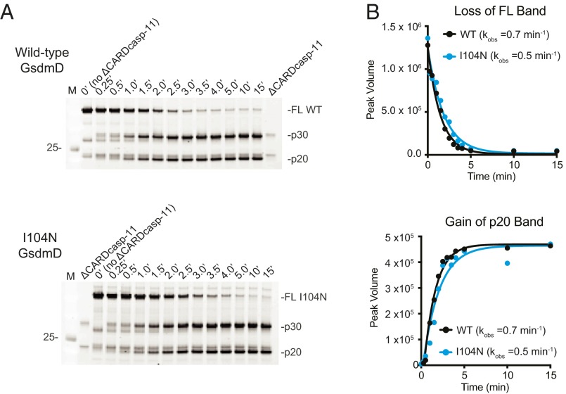
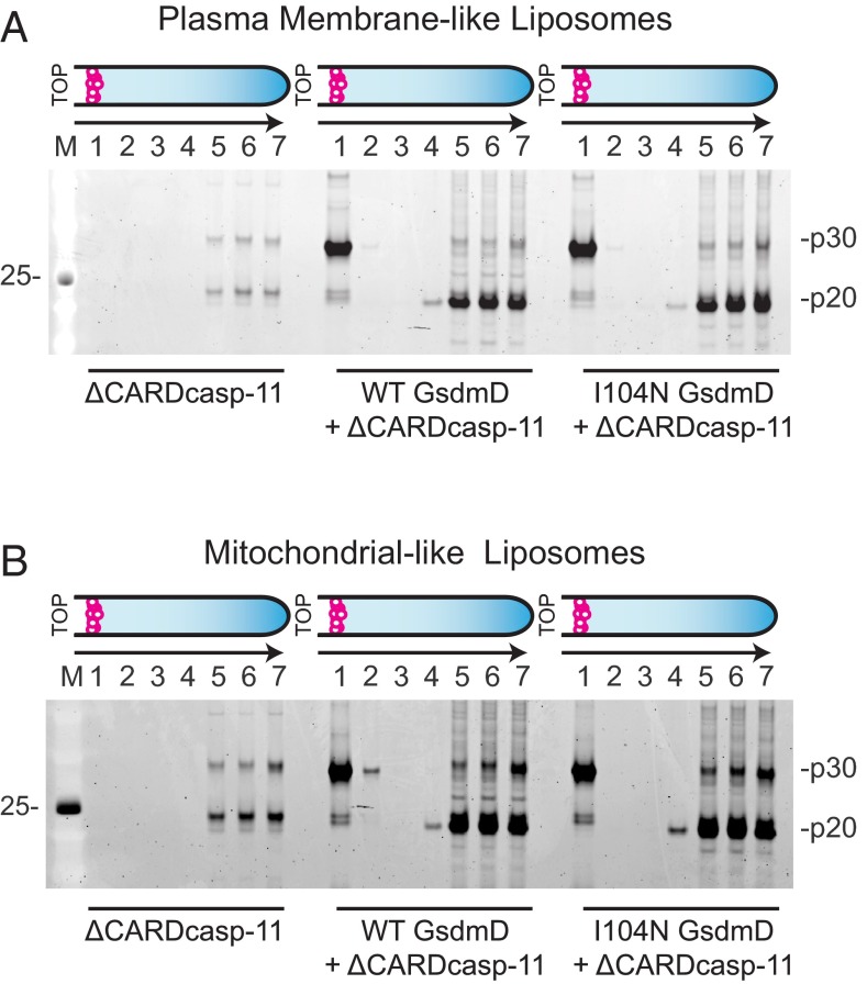
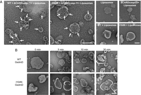
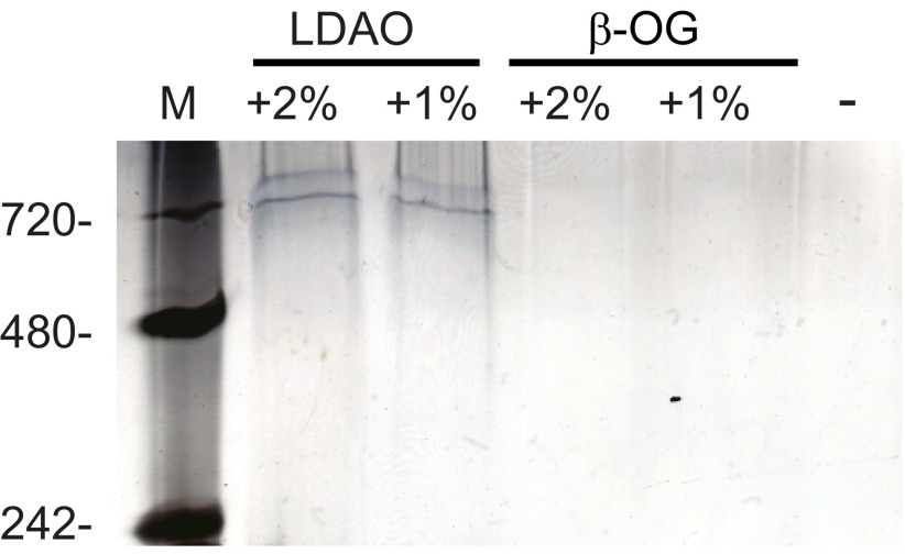
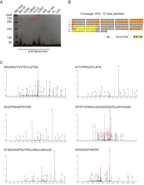
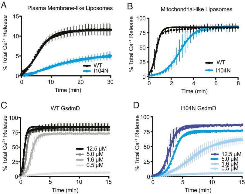
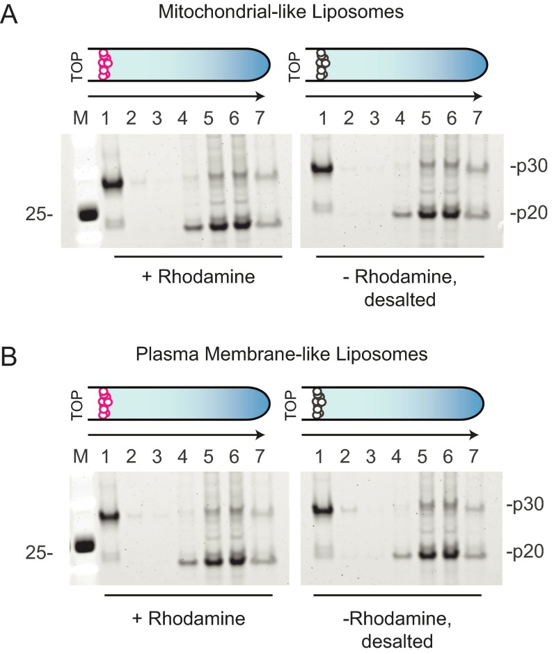
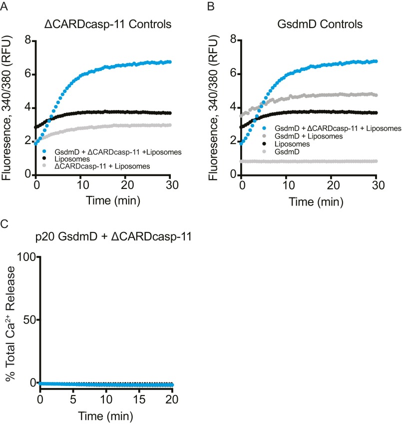
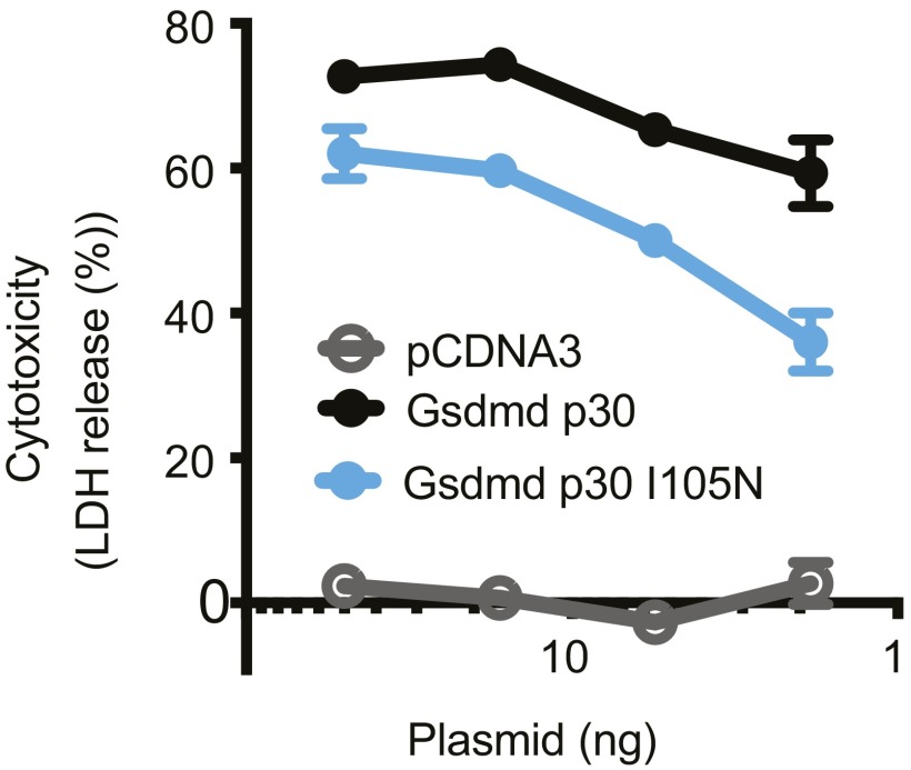
Gasdermin-D (GsdmD) is a critical mediator of innate immune defense because its cleavage by the inflammatory caspases 1, 4, 5, and 11 yields an N-terminal p30 fragment that induces pyroptosis, a death program important for the elimination of intracellular bacteria. Precisely how GsdmD p30 triggers pyroptosis has not been established. Here we show that human GsdmD p30 forms functional pores within membranes. When liberated from the corresponding C-terminal GsdmD p20 fragment in the presence of liposomes, GsdmD p30 localized to the lipid bilayer, whereas p20 remained in the aqueous environment. Within liposomes, p30 existed as higher-order oligomers and formed ring-like structures that were visualized by negative stain electron microscopy. These structures appeared within minutes of GsdmD cleavage and released Ca(2+) from preloaded liposomes. Consistent with GsdmD p30 favoring association with membranes, p30 was only detected in the membrane-containing fraction of immortalized macrophages after caspase-11 activation by lipopolysaccharide. We found that the mouse I105N/human I104N mutation, which has been shown to prevent macrophage pyroptosis, attenuated both cell killing by p30 in a 293T transient overexpression system and membrane permeabilization in vitro, suggesting that the mutants are actually hypomorphs, but must be above certain concentration to exhibit activity. Collectively, our data suggest that GsdmD p30 kills cells by forming pores that compromise the integrity of the cell membrane.

Keywords: GsdmD; caspase-11; pyroptosis.


Conflict of interest statement

Conflict of interest statement: R.A.A., A.E., A.G., P.S.L., N.K., C.C., V.M.D., and E.C.D. were employees of Genentech, Inc.

Figures

Fig. 1.
Fig. 1.
Processing and membrane partitioning of GsdmD. (A) SDS/PAGE of wild-type (WT) and I104N GsdmD with and without ΔCARDcasp-11 treatment (coomassie staining). Full-length (FL) and cleavage products, p20 and p30, are denoted. (B) WT GsdmD cleavage reactions were subject to sedimentation analysis to assess component solubilities. SDS/PAGE of input (I), supernatant (S), wash (W), and pellet (P) fractions are shown for different washing strategies (see SI Methods, SYPRO ruby staining). (C) Schematic of liposome flotation assay. A high-density medium was used to float the low-density liposomes and any associated proteins. Rhodamine-labeled liposomes are shown as magenta circles, and protein is shown as gray stars. (D and E) Liposome flotation assay fractions were evaluated by SDS/PAGE (SYPRO ruby staining) for reactions containing (D) plasma membrane-like and (E) mitochondrial-like liposomes. Lanes correspond to successive 100-μL layers starting at the top of the assay tube (Left to Right) and solubilized pellet (fraction 7). Liposome quantification of each layer (as measured by rhodamine fluorescence, normalized to the top layer) is depicted in the bar graphs above the gels. (F) Western blots of immortalized macrophages after stimulation with intracellular LPS. Cellular localization markers, GAPDH (glyceraldehyde 3-phosphate dehydrogenase), Na+/K+-ATPase (sodium-potassium adenosine triphosphatase), and VDAC (voltage-dependent ion channel), are shown below.
Fig. S1.
Fig. S1.
Wild-type (WT) GsdmD colocalization with plasma membrane-like liposomes lacking sphingomyelin. Mitochondrial-like and plasma membrane-like liposomes lacking sphingomyelin (Table S1) were incubated with wild-type GsdmD cleavage reactions. Liposomes were then separated using the flotation assay and analyzed by SDS/PAGE (SYPRO ruby staining). The GsdmD cleavage products p20 and p30 are shown. Liposome quantification of each coflotation layer is represented in a bar graph above the gel as rhodamine fluorescence, normalized to the top layer.
Fig. S2.
Fig. S2.
Western blots of bone marrow-derived macrophages from indicated mice. Wild-type (WT).
Fig. 2.
Fig. 2.
Kinetics of GsdmD cleavage by ΔCARDcasp-11. (A) SDS/PAGE analysis of wild-type (WT) and I104N GsdmD cleavage reaction timecourses (SYPRO ruby staining). (B) Quantification of full-length (FL) and p20 bands for WT (black) and I104N (blue) reactions. Single-exponential fits to the data were used to determine listed rates.
Fig. 3.
Fig. 3.
GsdmD colocalization with different membrane compositions. Rhodamine-labeled (A) plasma membrane-like and (B) mitochondrial-like liposomes were incubated with wild-type (WT) and I104N cleavage reactions or with ΔCARDcasp-11 alone. Liposomes were separated via flotation assay, and the resulting layers were analyzed by SDS/PAGE (SYPRO ruby staining). GsdmD p20 and p30 cleavage products are denoted.
Fig. 4.
Fig. 4.
Negative stain electron microscopy images of p30-bound liposomes. (A) Representative images of liposomes incubated with GsdmD cleavage reactions and control samples. Ring-shaped pore structures are highlighted with arrows. (B) Images from a timecourse of GsdmD cleavage reactions initiated by ΔCARDcasp-11. (Scale bar for all images, 100 nm.)
Fig. 5.
Fig. 5.
Blue Native-PAGE analysis of p30 colocalized to membranes. Liposome-bound p30 was treated with detergent and analyzed by BN-PAGE along with a no-detergent control. Bands were visualized by silver staining. LDAO (n-Dodecyl-N,N-Dimethylamine-N-Oxide) and β-OG (n-Octyl-β-Glucoside).
Fig. S3.
Fig. S3.
Detergent extraction of GsdmD p30 complex from liposomes. (A) Blue native page gel (coomassie staining) of p30 extraction from liposomes using the panel of detergents listed. The band identified as p30 is indicated by a red arrow. (B) Coverage map of GsdmD p30 sequence. (C) Representative tandem mass spectra confirming the 720-kDa band as GsdmD p30.
Fig. 6.
Fig. 6.
Calcium-loaded liposome release assays. Calcium release from (A) plasma membrane-like and (B) mitochondrial-like liposomes upon incubation with ΔCARDcasp-11 and full-length wild-type (WT) or I104N GsdmD (mean ± SD error bars; n = 6). Dilution series of (C) WT and (D) I104N GsdmD in calcium release assays from loaded mitochondrial-like liposomes (mean ± error bars; n = 3). All fits shown are standard logistic function.
Fig. S4.
Fig. S4.
Coflotation assay of desalted liposomes. (A) Mitochondrial-like and (B) plasma membrane-like liposomes were prepared ± rhodamine label. Liposomes without the rhodamine label were desalted via NAP-5 column. All liposomes were then incubated with GsdmD cleavage reactions, separated via flotation assay, and analyzed via SDS/PAGE (SYPRO ruby staining). Successive layers of the coflotation assay, as well as GsdmD cleavage products p20 and p30, are shown.
Fig. S5.
Fig. S5.
Cargo release assay background signal subtraction and controls. (A) ΔCARDcasp-11 background signal. The raw ratio of fluorescence from 340/380 nm excitation is shown for a GsdmD cleavage reaction in the presence of liposomes (blue), as well as the background signal for liposomes alone (black) and liposomes + ΔCARDcasp-11 (gray). Because the ΔCARDcasp-11 storage buffer contains imidazole that binds a fraction of the background calcium signal (compare liposomes alone to liposomes + ΔCARDcasp-11), a parallel ΔCARDcasp-11 + liposomes control assay was used for background subtraction of the data reported in Fig. 6. (B) GsdmD liposome release controls. The raw ratio of fluorescence from 340/380 nm excitation is shown for a GsdmD cleavage reaction in the presence of liposomes (blue), liposomes alone (black), GsdmD + liposomes (dark gray), and GsdmD alone (light gray). (C) GsdmD p20 liposome cargo release assay control. CaCl2-loaded mitochondrial-like liposomes were incubated with ΔCARDcasp-11 and GsdmD p20 and monitored for calcium release (mean ± error bars; n = 3).
Fig. 7.
Fig. 7.
Cytotoxicity of GsdmD p30 variants. Cytotoxicity, as measured by lactate dehydrogenase (LDH) release, at 24 h after transient transfection of HEK293T cells. Graph shows the mean ± SD of triplicate wells and is representative of three independent experiments.

References

    1. Cookson BT, Brennan MA. Pro-inflammatory programmed cell death. Trends Microbiol. 2001;9(3):113–114. - PubMed
    1. Lamkanfi M, Dixit VM. Mechanisms and functions of inflammasomes. Cell. 2014;157(5):1013–1022. - PubMed
    1. Miao EA, et al. Caspase-1-induced pyroptosis is an innate immune effector mechanism against intracellular bacteria. Nat Immunol. 2010;11(12):1136–1142. - PMC - PubMed
    1. Kayagaki N, et al. Non-canonical inflammasome activation targets caspase-11. Nature. 2011;479(7371):117–121. - PubMed
    1. Kayagaki N, et al. Noncanonical inflammasome activation by intracellular LPS independent of TLR4. Science. 2013;341(6151):1246–1249. - PubMed