. 2017 Mar;37(3):1120-1136.
doi: 10.1177/0271678X16655172. Epub 2016 Jul 20.

Proteomic differences in brain vessels of Alzheimer's disease mice: Normalization by PPARγ agonist pioglitazone

Affiliations

Proteomic differences in brain vessels of Alzheimer's disease mice: Normalization by PPARγ agonist pioglitazone

AmanPreet Badhwar et al. J Cereb Blood Flow Metab. 2017 Mar.

Abstract

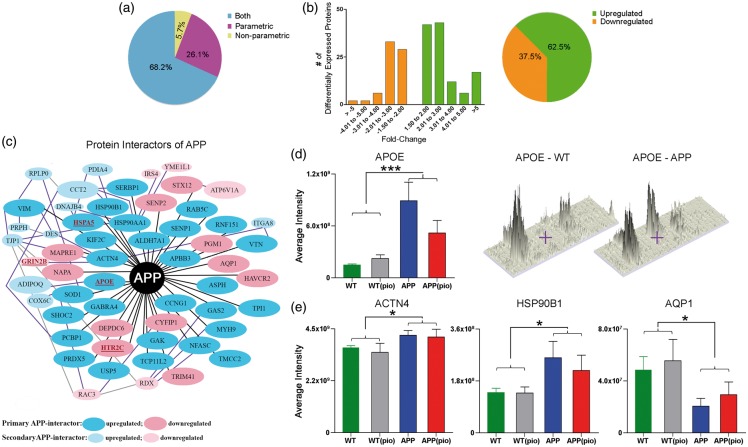
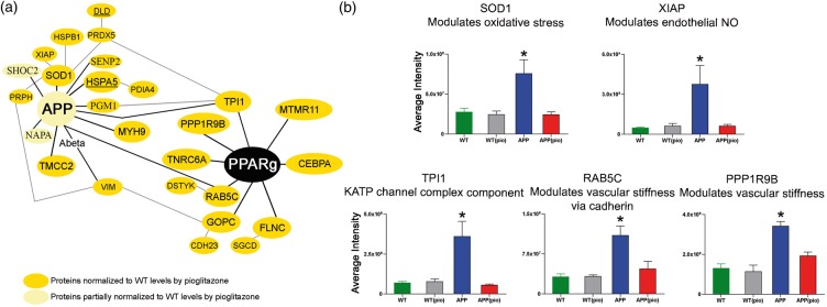
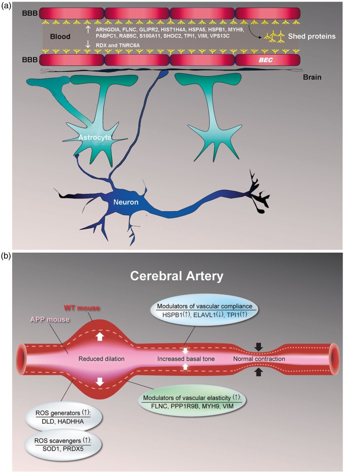
Cerebrovascular insufficiency appears years prior to clinical symptoms in Alzheimer's disease. The soluble, highly toxic amyloid-β species, generated from the amyloidogenic processing of amyloid precursor protein, are known instigators of the chronic cerebrovascular insufficiency observed in both Alzheimer's disease patients and transgenic mouse models. We have previously demonstrated that pioglitazone potently reverses cerebrovascular impairments in a mouse model of Alzheimer's disease overexpressing amyloid-β. In this study, we sought to characterize the effects of amyloid-β overproduction on the cerebrovascular proteome; determine how pioglitazone treatment affected the altered proteome; and analyze the relationship between normalized protein levels and recovery of cerebrovascular function. Three-month-old wildtype and amyloid precursor protein mice were treated with pioglitazone- (20 mg/kg/day, 14 weeks) or control-diet. Cerebral arteries were surgically isolated, and extracted proteins analyzed by gel-free and gel-based mass spectrometry. 193 cerebrovascular proteins were abnormally expressed in amyloid precursor protein mice. Pioglitazone treatment rescued a third of these proteins, mainly those associated with oxidative stress, promotion of cerebrovascular vasocontractile tone, and vascular compliance. Our results demonstrate that amyloid-β overproduction perturbs the cerebrovascular proteome. Recovery of cerebrovascular function with pioglitazone is associated with normalized levels of key proteins in brain vessel function, suggesting that pioglitazone-responsive cerebrovascular proteins could be early biomarkers of Alzheimer's disease.

Keywords: Amyloid peptide; cerebral artery; oxidative stress; proliferator-activated receptor gamma; vascular biomarkers.


Figures

Figure 1.
Figure 1.
Quantification of APP protein levels in cerebral arteries. (a) Alignment of the detected APP peptide (LVFFAEDVGSNK) in relation to the full-length APP protein. Also, indicated are the cleavage sites for the α, β, and γ secretases, as well as the AD-associated Swedish (KM) and Indiana (V) mutations. (b) Peptide intensity distribution of WT and APP mice as visualized using MSight. For both genotypes, biological replicate 1, each consisting of vessels extracted from three different mice is shown. The shaded grey area and/or the purple cross hair represent APP protein level. (c) As expected, APP mice (blue) display significantly elevated cerebrovascular APP protein levels relative to WT controls (green). Pioglitazone therapy moderately rescued APP protein levels in treated APP (red), but had no effect on WT (grey) mice. Error bars denote standard error or SEM and ⋆⋆ p < 0.01. PIO: pioglitazone.
Figure 2.
Figure 2.
Proteins differentially expressed in brain vessels of APP mice. (a) Distribution (in percentage) of proteins identified with either parametric or non-parametric Student t-test or both statistical analyses. (b) Fold-change distribution of differentially expressed proteins along with the percentage of upregulated or downregulated proteins in the APP vasculature. (c) Protein interactors of APP or proteins known to directly (black line) or indirectly (secondary: purple line and tertiary: grey line interactions only) interact with APP were identified using the STRING, IntAct, and in-house, and/or PubMed reference library. Note that only a few indirectly interacting proteins have been shown. Proteins associated with increased AD risk in the AlzGene database and/or by querying the PubMed reference library are identified in red and underlined (e.g. APOE). Average (d) APOE, and (e) HSP90B1, AQP1, and ACTN4 peptide/s intensities for WT (green), WT(pio) (grey), APP(blue), and APP(pio) (red). Error bars denote standard error or SEM, ⋆ p < 0.05 and ⋆⋆⋆ p < 0.001 denote significant genotype effect using two-way ANOVA. PIO: pioglitazone.
Figure 3.
Figure 3.
Pioglitazone-recovered proteins in the APP vasculature. (a) Pioglitazone-rescued protein that (i) directly or indirectly interact with APP, and/or (ii) harbor known PPARγ-PPRE/PACM binding sites in their genomic sequences. (b) Average SOD1, XIAP, TPI1, RAB5C, and PPP1R9B peptide intensities for WT (green), WT(pio) (grey), APP (blue), and APP(pio) (red) mice. Note: The function listed for RAB5C is probable, but not confirmed. Error bars denote standard error or SEM and ⋆ p < 0.05 denote significant interaction effect using two-way ANOVA. NO: nitric oxide; PIO: pioglitazone.
Figure 4.
Figure 4.
Cerebrovascular functional recovery by pioglitazone in APP mice. (a) 16 cerebrovascular shed proteins (or proteins present in circulating BEC microvesicles) constituting 27% of pioglitazone-rescued proteins were identified. Fourteen of these shed proteins were upregulated (↑) in APP mice and normalized by pioglitazone: ARHGDIA, FLNC, GLIPR2, HIST1H4A, HSPA5, HSPB1, MYH9, PABPC1, RAB5C, S100A11, SHOC2, TPI1, VIM, and VPS13C, whereas two were downregulated (↓) in APP mice and normalized by treatment: RDX and TNRC6A. These proteins could serve as surrogates to indicate efficacy and/or select responders in pioglitazone clinical trials. (b) APP overexpression and consequent Aβ increase in APP mice alter levels of proteins involved in mediating (i) oxidative stress (both ROS generators and scavengers), (ii) vascular elasticity and compliance, and (iii) increased basal tone, which then promotes impaired vasodilation. Pioglitazone treatment normalizes levels of these proteins and recovers cerebrovascular function. Of the proteins shown, FLNC, HSPB1, MYH9, TPI1, and VIM are shed proteins. Symbols: ↑, up and ↓, down.

References

    1. Ruitenberg A, den Heijer T, Bakker SL, et al. Cerebral hypoperfusion and clinical onset of dementia: the Rotterdam Study. Ann Neurol 2005; 57: 789–794. - PubMed
    1. Nicolakakis N, Hamel E. Neurovascular function in Alzheimer's disease patients and experimental models. J Cereb Blood Flow Metab 2011; 31: 1354–1370. - PMC - PubMed
    1. Wierenga CE, Hays CC, Zlatar ZZ. Cerebral blood flow measured by arterial spin labeling MRI as a preclinical marker of Alzheimer's disease. J Alzheimers Dis 2014; 42: S411–S419. - PMC - PubMed
    1. Kalaria RN. Cerebral vessels in ageing and Alzheimer's disease. Pharmacol Ther 1996; 72: 193–214. - PubMed
    1. Farkas E, Luiten PG. Cerebral microvascular pathology in aging and Alzheimer's disease. Prog Neurobiol 2001; 64: 575–611. - PubMed

MeSH terms

LinkOut - more resources