Review
. 2016 Jun 23;14(1):52.
doi: 10.1186/s12951-016-0195-8.

Application prospective of nanoprobes with MRI and FI dual-modality imaging on breast cancer stem cells in tumor

Affiliations
Review

Application prospective of nanoprobes with MRI and FI dual-modality imaging on breast cancer stem cells in tumor

Hetao Chen et al. J Nanobiotechnology. .

Abstract

Breast cancer (BC) is a serious disease to threat lives of women. Numerous studies have proved that BC originates from cancer stem cells (CSCs). But at present, no one approach can quickly and simply identify breast cancer stem cells (BCSCs) in solid tumor. Nanotechnology is probably able to realize this goal. But in study process, scientists find it seems that nanomaterials with one modality, such as magnetic resonance imaging (MRI) or fluorescence imaging (FI), have their own advantages and drawbacks. They cannot meet practical requirements in clinic. The nanoprobe combined MRI with FI modality is a promising tool to accurately detect desired cells with low amount in tissue. In this work, we briefly describe the MRI and FI development history, analyze advantages and disadvantages of nanomaterials with single modality in cancer cell detection. Then the application development of nanomaterials with dual-modality in cancer field is discussed. Finally, the obstacles and prospective of dual-modal nanoparticles in detection field of BCSCs are also pointed out in order to speed up clinical applications of nanoprobes.

Keywords: Breast cancer; Cancer stem cells; Fluorescence; Imaging; Magnetic resonance.


Figures
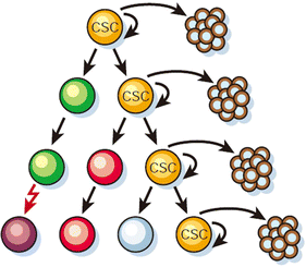
Fig. 1
Fig. 1
Most cancer cells have only limited proliferative potential, only CSCs (yellow) have the ability to proliferate extensively and form new tumors [5] [Adapted by permission from Macmillan Publishers Ltd: [Nature] (Ref.5), copyright (2001)]
Fig. 2
Fig. 2
Emission colors of QDs excited by UV
Fig. 3
Fig. 3
QD-based double-color in situ fluorescent imaging for Ki67 andHER2 in BC. QD-based double-color images for Ki67 andHER2, Ki67was expressed as clear red fluorescence, HER2 as bright green fluorescence (a); the spectral images of Ki67 and HER2 co-expressions were obtained by CRi Nuance multispectral imaging system, which could unmix the images into single color images (b); the single red fluorescent image representing Ki67 at the emitting wavelength of 655 nm (c); and the single green fluorescent representing HER2 at the emitting wavelength of 525 nm (d). 200× , scale bar = 50 μm [28]
Fig. 4
Fig. 4
a In vivo simultaneous imaging of multicolor QD-encoded microbeads injected into a live mouse (b) Molecular targeting and in vivo imaging of a prostate tumor in mouse using a QD–antibody conjugate (red) [32]
Fig. 5
Fig. 5
1.5-T MRI turbo-spin-echo-T2-weighted (5500/100) dynamic imaging of human MDA- MB-231 breast cancer xenografts. af Injection of γ-Fe2O3@DMSA NPs showed that the tumor (thick white arrows) signal intensity decreased at 12 h which returned to basal levels by 24 h. gl Injection of γ-Fe2O3@DMSA-DG NPs showed that the tumor signal intensity decreased between 12 and 48 h, with the most hypointensity observed at 24 h. The signal intensity in the liver (thin white arrows) significantly decreased after injection of γ-Fe2O3@DMSA NPs or γ-Fe2O3@DMSA-DG NPs [48]
Fig. 6
Fig. 6
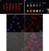
In vitro multimodal imaging with nanoprobes. (a) Fluorescence image of cell pellets and (b) MR (upper) and its color mapped (lower) images of dispersed cells in agarose. (c) Confocal laser scanning microscopic images [57]. Reprinted with permission from (J Am Chem Soc. 2010; 132: 552–557). Copyright (2010) American Chemical Society
Fig. 7
Fig. 7
Upconversion fluorescence images of a mouse injected with multifunctional nanoparticles-labeled mMSCs taken right after injection (a) and 6 h after injection (b) in the presence of a magnetic field. (c) In vivo MR image of the same mouse in (b) [68]
Fig. 8
Fig. 8
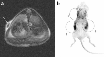
In vivo MR imaging (a) and fluorescence imaging (b) of nude mouse after subcutaneous injection of labeled and unlabeled human mesenchymal stem cells with MNP@SiO2(RITC)-PEG (1 = 1 × 105 unlabeled human mesenchymal stem cells for control, 2 = 1 × 106 labeled human mesenchymal stem cells, 3 = 1 × 105 labeled human mesenchymal stem cells). Labeled human mesenchymal stem cells are clearly seen as dark dot (arrow) in subcutaneous layer of nude mouse on axial scan of fast spin echo sequence. Fluorescent signal of subcutaneously injected human mesenchymal stem cells is detected at injection sites of labeled cells. Injection sites of unlabeled cells shows autofluorescence-induced artifact [69]

References

    1. Siegel RL, Miller K, Jemal AJ. Cancer statistics, 2016. CA-Cancer J Clin. 2016;65:7–30. doi: 10.3322/caac.21332. - DOI - PubMed
    1. Dennis JS, Gary MC, Steven GW, Wendy JL, Axel U, McGuire WL. Human breast cancer: correlation of relapse and survival with amplification of the HER-2/neu oncogene. Science. 1987;235:177–182. doi: 10.1126/science.3798106. - DOI - PubMed
    1. Lin NU, Claus E, Sohl J, Razzak AR, Arnaout A, Winer EP. Sites of distant recurrence and clinical outcomes in patients with metastatic triple-negative breast cancer. Cancer. 2008;113:2638–2645. doi: 10.1002/cncr.23930. - DOI - PMC - PubMed
    1. Early Breast Cancer Trialist`s Collaborative Group: effects of chemotherapy and hormonal therapy for early breast cancer on recurrence and 15-year survival: an overview of the randomised trials. Lancet. 2005;365:1687–1717. doi: 10.1016/S0140-6736(05)66544-0. - DOI - PubMed
    1. Reya T, Morrison SJ, Clarke MF, Weissman IL. Stem cells, cancer, and cancer stem cells. Nature. 2001;414:105–111. doi: 10.1038/35102167. - DOI - PubMed

MeSH terms