. 2016 Jun 24:6:28604.
doi: 10.1038/srep28604.

SAS6-like protein in Plasmodium indicates that conoid-associated apical complex proteins persist in invasive stages within the mosquito vector

Affiliations

SAS6-like protein in Plasmodium indicates that conoid-associated apical complex proteins persist in invasive stages within the mosquito vector

Richard J Wall et al. Sci Rep. .

Abstract

The SAS6-like (SAS6L) protein, a truncated paralogue of the ubiquitous basal body/centriole protein SAS6, has been characterised recently as a flagellum protein in trypanosomatids, but associated with the conoid in apicomplexan Toxoplasma. The conoid has been suggested to derive from flagella parts, but is thought to have been lost from some apicomplexans including the malaria-causing genus Plasmodium. Presence of SAS6L in Plasmodium, therefore, suggested a possible role in flagella assembly in male gametes, the only flagellated stage. Here, we have studied the expression and role of SAS6L throughout the Plasmodium life cycle using the rodent malaria model P. berghei. Contrary to a hypothesised role in flagella, SAS6L was absent during gamete flagellum formation. Instead, SAS6L was restricted to the apical complex in ookinetes and sporozoites, the extracellular invasive stages that develop within the mosquito vector. In these stages SAS6L forms an apical ring, as we show is also the case in Toxoplasma tachyzoites. The SAS6L ring was not apparent in blood-stage invasive merozoites, indicating that the apical complex is differentiated between the different invasive forms. Overall this study indicates that a conoid-associated apical complex protein and ring structure is persistent in Plasmodium in a stage-specific manner.


Figures

Figure 1
Figure 1. SAS6L gene transcript and protein expression throughout the life cycle in P.berghei.
(a) Transcription of sas6l analysed by qRT-PCR, normalised against two endogenous control genes, hsp70 and arginine-tRNA synthetase (Pfaffl method). Each bar is the mean of three biological replicates ± SEM. All asexual blood stages: AS; schizonts: Sch; non-activated gametocytes: NAG; activated gametocytes: AG; ookinete: Ook; 14 dpi oocysts/sporozoites: Spor. (b) Live fluorescence detection of SAS6L-GFP in blood-stage schizont, female and male gametes, male gamete with flagella, zygote, ookinetes, oocyst (14 dpi) and sporozoite (21 dpi). 13.1, a cy3-conjugated antibody which recognises P28 on the surface of activated female, zygote, and ookinete (red) was used with the sexual stages. Nuclei were detected using Hoechst 33342 (blue), and the cells were displayed by differential interference contrast (DIC). Merge is the composite of Hoechst, GFP and 13.1 signals. Insets show magnified view of SAS6L-GFP dots. White arrow points to single flagellated male gamete. Scale bars = 5 μm.
Figure 2
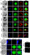
Figure 2. Developmental profile of SAS6L in ookinetes and sporozoites.
(a) Localisation of SAS6L-GFP throughout the different stages of ookinete development (from early zygote to ookinete stages). 13.1 labels the surface (red) of the sexual stages as for Fig. 1. Nuclei were detected using Hoechst 33342 (blue), and the cells were displayed by differential interference contrast (DIC). Merge is the composite of Hoechst, GFP and 13.1. (b) Localisation of SAS6L-GFP throughout the mosquito development (from 7 dpi to 21 dpi). Merge is the composite of Hoechst and GFP. (c) Localisation of SAS6L-GFP in isolated 14 and 21 dpi sporozoites. Scale bars = 5 μm.
Figure 3
Figure 3. 3D-SIM super-resolution images of SAS6L shows a ring structure at the apical end of the ookinete and sporozoite.
Images of fixed zygotes, ookinetes, and salivary gland sporozoite at 21 days post-infection expressing SAS6L-GFP (green), co-stained with DAPI (blue), 13.1 (red: zygotes/ookinetes) or CSP (red: sporozoites). Images were selected for the orientation of the apical end of the cell facing the viewer and displaying the ring. Major scale bars = 5 μm. Insets show magnified view of SAS6L ring structures with scale bars = 500 nm.
Figure 4
Figure 4. SAS6L location and behaviour throughout the tachyzoite cell cycle in Toxoplasma gondii.
T. gondii expressing endogenous SAS6L with C-terminal fusion to HA-APEX fixed in host vacuoles and stained for HA (green) and either IMC1 (red, a and c) or centrin (red, b) (a) 3D-SIM super-resolution images with cell apices facing the viewer showing SAS6L rings (arrows, insets) and apices in profile showing SAS6L flush with the apical IMC. (b) Widefield images of parasites at different points of the cell cycle indicated by centrosome number and position (stained for centrin): interphase with single centrosome per cell (i); newly divided centrosomes close together (ii, upper vacuole); divided centrosomes migrated further apart (ii, lower vacuole; and iii); cells with nascent apical complexes (iv, arrowheads). (c) Widefield images of parasites showing daughter pellicle formation (stained for IMC1): before daughter pellicle formation (i and ii, upper vacuoles); small pellicle cups (i and ii, lower vacuoles; iii, upper vacuole); mid pellicle development (iii, lower vacuole). Scale bars = (a) 5 μm and 500 nm (inset); and (b,c) 10 μm.
Figure 5
Figure 5. Gene deletion shows that sas6l is not essential in parasite development and transmission.
(a) Microgametogenesis of ∆sas6l in comparison to WT measured as the number of exflagellation centres per field. Means ± SEM are shown. n = 22 from three independent experiments (cl. 2 = 2 experiments; cl. 3 = 1 experiment). (b) Ookinete conversion as a percentage in ∆sas6l and WT lines. Ookinetes were identified using the marker 13.1 and were defined as those cells that successfully differentiated into elongated ‘banana shaped’ ookinetes. Bar is the mean ± SEM. n = 4 (cl. 2 = 3; cl. 3 = 1) independent experiments. (c) Oocysts per mosquito gut (14 days post-infection; bar = arithmetic mean ± SEM; n = 38 from three independent experiments. (cl. 2 = 2 experiments; cl. 3 = 1 experiment) of Δsas6l or WT parasite-infected mosquitoes. An infection rate of >80% was observed for both Δsas6l and WT parasites. (d) Sporozoites per mosquito for 14 and 21 dpi and bite-back experiments indicating successful transmission of both Δsas6l and WT parasites from mosquito to mouse. Infected mosquitoes were allowed to feed and the mice were monitored for blood stage parasitaemia (days post infection, dpi) indicative of successful liver and blood stage infection. Example of two independent experiments (cl. 2 and cl. 3) are shown. (e) Relative expression of sas6, sas6l, isp1, isp3, nek2, nek4, ppkl RNA in Δsas6l parasites and (f), sas6l RNA level in Δdozi mutant parasites compared to WT controls (∆∆Ct method). Mean ± SEM, n = 3 biological replicates from at least two independent experiments. One representative experiment is shown.

Similar articles

Cited by

References

    1. Leidel S., Delattre M., Cerutti L., Baumer K. & Gonczy P. SAS-6 defines a protein family required for centrosome duplication in C. elegans and in human cells. Nat. Cell Biol. 7, 115–125 (2005). - PubMed
    1. Nakazawa Y., Hiraki M., Kamiya R. & Hirono M. SAS-6 is a cartwheel protein that establishes the 9-fold symmetry of the centriole. Curr. Biol. 17, 2169–2174 (2007). - PubMed
    1. de Leon J. C. et al.. A SAS-6-Like protein suggests that the Toxoplasma conoid complex evolved from flagellar components. Eukaryot. Cell 12, 1009–1019 (2013). - PMC - PubMed
    1. van Breugel M., Wilcken R., McLaughlin S. H., Rutherford T. J. & Johnson C. M. Structure of the SAS-6 cartwheel hub from Leishmania major. eLife 3 (2014). - PMC - PubMed
    1. Marques S. R. et al.. An essential role of the basal body protein SAS-6 in Plasmodium male gamete development and malaria transmission. Cell. Microbiol. 17, 191–206 (2015). - PMC - PubMed

Publication types

Substances

LinkOut - more resources