. 2016 Aug 19;291(34):17727-42.
doi: 10.1074/jbc.M116.735258. Epub 2016 Jun 23.

Asn347 Glycosylation of Corticosteroid-binding Globulin Fine-tunes the Host Immune Response by Modulating Proteolysis by Pseudomonas aeruginosa and Neutrophil Elastase

Affiliations

Asn347 Glycosylation of Corticosteroid-binding Globulin Fine-tunes the Host Immune Response by Modulating Proteolysis by Pseudomonas aeruginosa and Neutrophil Elastase

Zeynep Sumer-Bayraktar et al. J Biol Chem. .

Abstract

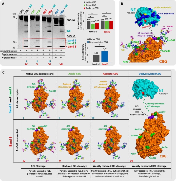
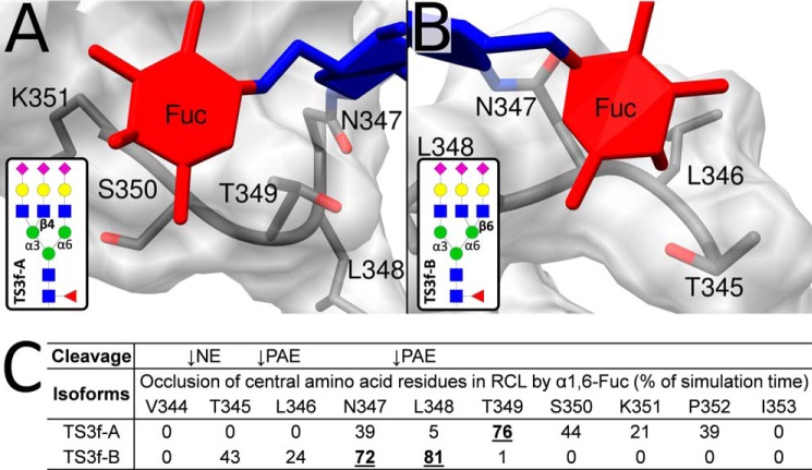
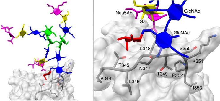
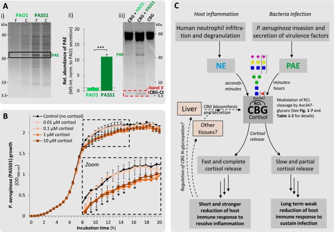
Corticosteroid-binding globulin (CBG) delivers anti-inflammatory cortisol to inflamed tissues upon elastase-based proteolysis of the exposed reactive center loop (RCL). However, the molecular mechanisms that regulate the RCL proteolysis by co-existing host and bacterial elastases in inflamed/infected tissues remain unknown. We document that RCL-localized Asn(347) glycosylation fine-tunes the RCL cleavage rate by human neutrophil elastase (NE) and Pseudomonas aeruginosa elastase (PAE) by different mechanisms. NE- and PAE-generated fragments of native and exoglycosidase-treated blood-derived CBG of healthy individuals were monitored by gel electrophoresis and LC-MS/MS to determine the cleavage site(s) and Asn(347) glycosylation as a function of digestion time. The site-specific (Val(344)-Thr(345)) and rapid (seconds to minutes) NE-based RCL proteolysis was significantly antagonized by several volume-enhancing Asn(347) glycan features (i.e. occupancy, triantennary GlcNAc branching, and α1,6-fucosylation) and augmented by Asn(347) NeuAc-type sialylation (all p < 0.05). In contrast, the inefficient (minutes to hours) PAE-based RCL cleavage, which occurred equally well at Thr(345)-Leu(346) and Asn(347)-Leu(348), was abolished by the presence of Asn(347) glycosylation but was enhanced by sialoglycans on neighboring CBG N-sites. Molecular dynamics simulations of various Asn(347) glycoforms of uncleaved CBG indicated that multiple Asn(347) glycan features are modulating the RCL digestion efficiencies by NE/PAE. Finally, high concentrations of cortisol showed weak bacteriostatic effects toward virulent P. aeruginosa, which may explain the low RCL potency of the abundantly secreted PAE during host infection. In conclusion, site-specific CBG N-glycosylation regulates the bioavailability of cortisol in inflamed environments by fine-tuning the RCL proteolysis by endogenous and exogenous elastases. This study offers new molecular insight into host- and pathogen-based manipulation of the human immune system.

Keywords: P. aeruginosa elastase; Pseudomonas aeruginosa (P. aeruginosa); corticosteroid-binding globulin; cortisol; glycoprotein; glycosylation; molecular dynamics; neutrophil elastase; proteolysis; reactive center loop.


Figures

FIGURE 1.
FIGURE 1.
Highly specific NE-based RCL cleavage proximal to the Asn347 glycosylation of CBG. A, generation of the two complementary sets of CBG fragments upon NE (light blue) cleavage (i.e. CBG-Ct (band 1, 12 kDa (blue); band 2, 11 kDa (green); band 3, 5 kDa (red)) and CBG-Nt (50–55 kDa) fragments as evaluated by their differential migration on SDS-PAGE. Broken line, non-neighboring gel lanes from the same gel. B, MS-based profiles of the NE-generated Asn347-glycosylated CBG-Ct fragments after trypsin digestion (left). The structures of the glycopeptides were confirmed using CID-MS/MS as exemplified by the fragmentation of a TS3 Asn347 glycopeptide (right) and our previous glycoprofiling of native CBG (36). C, band 3 contained the Asn347-non-glycosylated variant of the CBG-Ct fragment as confirmed by the identification of the non-modified peptide 345TLNLTSKPIILR356. D, overview of the nomenclature and structures of the six most abundant glycans harboring Asn347 (i.e. BS2, BS2f (biantennary (B) glycans in light orange); TS2, TS2f, TS3, and TS3f (triantennary (T) glycans in dark orange)). f, α1,6-linked (core) fucosylation; S, number of NeuAc residues. E, bands 1 and 2 contained primarily tri- and biantennary Asn347 glycans, respectively. F, sequence of a selected region of the exposed RCL of CBG. Blue and black arrows indicate the identified cleavage sites of NE and trypsin, respectively.
FIGURE 2.
FIGURE 2.
The rapid NE-based RCL cleavage of CBG is affected by the Asn347 glycan occupancy, core fucosylation, and outer antennary GlcNAc branching. The relative levels of Asn347 glycan site occupancy (%) (A), core fucosylation (%) (B), and outer antennary GlcNAc branching (measured as the level of triantennary glycans) (%) (C) were measured on the tryptic peptide from the CBG-Ct fragment (345TLNLTSKPIILR356) as a function of NE-based digestion time (0–150 s) of CBG. See Fig. 3A for an extended NE-based incubation of CBG (i.e. 10 min and 4 h). Asn347-specific values of native human CBG based on previously reported data (36) are shown for the three studied glycan features (dotted lines). All data points are plotted as mean ± S.E. (error bars) (n = 3) and based on relative MS abundances except in A, where the occupancies are based the relative gel band intensities (see inset for the 5–25 s gel). The involvement of the three Asn347 glycan features was statistically tested by grouping the early (5–25 s, light orange) and the medium (30–150 s) digestion time points. *, p < 0.05; ***, p < 0.001. D, schematic illustration of the local molecular environment around Asn347 and the three glycan features affecting the NE-based RCL cleavage of CBG at Val344-Thr345.
FIGURE 3.
FIGURE 3.
Glycan occupancy of Asn347 abolishes the PAE-induced RCL cleavage. A, unlike NE-digested (light blue) CBG (two penultimate left lanes), crudely isolated and pure PAE (green) generated two relatively homogeneous complementary CBG fragments (i.e. a single CBG-Ct (band 3, 5 kDa) and a CBG-Nt (50–55 kDa) fragment under extended incubation (4 and 16 h, right lanes) as evaluated by SDS-PAGE. Undigested native CBG is shown in the far left lane. Broken line, non-neighboring gel lanes from the same gel. Solid line, gel lanes from different gels. B, amino acid residues of the exposed RCL of CBG in the vicinity of the observed PAE cleavage sites (green arrows). The CBG-Nt (gold) and CBG-Ct (brown) peptides covering the Asn347 glycosylation site are shown. C, the presence of 346LNLTSKPIILR356 and 348LTSKPIILR356 (data for the latter not shown) in an Asn347-non-glycosylated form (top EIC, left) and the absence of their corresponding Asn347-glycosylated forms (bottom EIC, left; as exemplified by the absence of an EIC signal for the TS3 Asn347 glycopeptide) in the CBG-Ct fragment was confirmed using CID-MS/MS (right). D, peptides of the complementary CBG-Nt fragments (i.e. 327AVLQLNEEGVDTAGSTGVTLN347 and 327AVLQLNEEGVDTAGSTGVT345 (data for the latter not shown) were also exclusively identified in their non-glycosylated state by LC-MS/MS. No EIC signals were detected in any charge states for any glycoforms of either the CBG-Nt or the CBG-Ct peptides. *, CBG-Nt fragments occurring after extended digestion of CBG with NE that were not further investigated in this study. ‡, crude PAE migrated around 33 kDa as validated by LC-MS/MS analysis, whereas the commercial form of PAE (“pure PAE”) was observed in separate experiments to co-migrate with human CBG around 55–60 kDa (data not shown).
FIGURE 4.
FIGURE 4.
Sialylation of the non-Asn347 glycans is beneficial for PAE-based RCL cleavage. A, incubations of (from left) undigested native CBG, undigested asialo-CBG, pure PAE-digested native CBG, pure PAE-digested asialo-CBG, undigested agalacto-CBG, pure PAE-digested agalacto-CBG, undigested deglycosylated CBG, and pure PAE-digested deglycosylated CBG. Shown is a comparison of the relative gel band intensities of band 3 (red box). Broken line, non-neighboring gel lanes from the same gel. Solid line, gel lanes from different gels. B, three-dimensional structure of PAE (PDB code 1EZM) and RCL-uncleaved CBG (homology model based on uncleaved human TBG, PDB code 2CEO). CBG is presented in a glycoform that allows RCL cleavage by PAE (i.e. all glycosylation sites are occupied except for Asn347). Color coding of monosaccharide residues is in accordance with the established nomenclature by the Consortium for Functional Glycomics (79). NeuAc residues are in purple, galactose residues are in yellow, GlcNAc residues are in blue, fucose residues are in red, and mannose residues are in green. Color coding of the polypeptide chains is as follows. The PAE cleavage sites (Thr345-Leu346 and Asn347-Leu348) on the RCL of CBG are in purple, the basic amino acid residues of PAE are in blue, acidic amino acid residues of PAE are in yellow, the amino acid residues in the active site of PAE are in red, and the Asn glycosylated sites of interest of CBG are in green. The proposed electrostatic interactions and protein docking are schematically illustrated with broken lines. C, proposed interactions of NE and glycosylated variants of CBG, which may explain the differential RCL cleavage efficiencies observed in A; same color coding as in B. **, p < 0.01; ***, p < 0.001; ns, not significant. Data points are represented as mean ± S.E. (error bars), n = 3.
FIGURE 5.
FIGURE 5.
Sialylation of Asn347 glycans, not of glycans from other CBG sites, is beneficial for NE-based RCL cleavage. A, incubations of (from left) undigested native CBG (black), NE-digested native CBG, undigested asialo-CBG (green), NE-digested asialo-CBG, undigested agalacto-CBG (red), NE-digested agalacto-CBG, undigested deglycosylated CBG (blue), and NE-digested deglycosylated CBG. Comparisons of the relative gel band intensities (red box) were performed. Broken line, non-neighboring gel lanes from the same gel. B, the three-dimensional structures of NE (PDB code 3Q77) and RCL uncleaved CBG (homology model based on uncleaved human TBG, PDB code 2CEO). CBG is presented with its most abundant glycoforms of the three sites located in proximity to the RCL (i.e. Asn347 displays TS3, and Asn154 and Asn238 both display BS2). The color coding of the monosaccharide residues and amino acid residues is generally as described in the legend to Fig. 4, the NE cleavage site (Val344-Thr345) on the RCL of CBG is in purple. The proposed electrostatic interactions and protein docking to the RCL of CBG are schematically illustrated with broken lines. C, proposed interactions of NE and the glycosylated variants of CBG, which may explain the differential RCL cleavage observed in Fig. 5A. *, p < 0.05; ns, not significant. Data points are represented as mean ± S.E. (error bars), n = 3.
FIGURE 6.
FIGURE 6.
MD simulations indicate that core fucosylation of Asn347 glycans occludes central amino acid residues of RCL. The α1,6-linked core fucose of the TS3f-A (Man 3′-arm GlcNAc branching) (A) and TS3f-B (Man 6′-arm GlcNAc branching) (B) isomers formed prolonged interactions with two different regions of the RCL. C, the interactions generated by the TS3f-B isomer resulted in significant occlusion of both the NE and PAE proteolytic sites as measured by the reduced solvent accessibility of the Thr345-Leu348 region generated by the core fucose residue over the simulation time. In contrast, the core fucose-centric interactions generated by the supposedly more abundant TS3f-A isomer influenced only the PAE digestion sites by occluding the Asn347–Pro352 region. Amino acid residues occluded for >50% of the simulation time are in boldface type and underlined. See Fig. 7 and Table 2 for the RCL occlusions provided by the entire Asn347 glycan moieties, not just their core fucose residues. See the legend to Fig. 1 and supplemental Table S1 for the nomenclature used for the Asn347 glycoforms.
FIGURE 7.
FIGURE 7.
MD simulations suggest that Asn347 glycans form sustained interactions to the RCL backbone of CBG that occlude primarily the PAE digestion sites. Shown is an example of Asn347 glycan-CBG protein surface interaction as demonstrated by the prolonged contact of the chitobiose core of TS3f-B to the PAE digestion sites of the RCL (left), in particular the Asn347–Lys351 region; see enlarged region (right) for details. See also Table 2 for details on the reduction of solvent accessibilities of TS3f-B and other Asn347 glycoforms over the MD simulation time. See the legend to Fig. 1 and supplemental Table S1 for the nomenclature used for the Asn347 glycoforms.
FIGURE 8.
FIGURE 8.
High PAE secretion and cortisol concentration-dependent growth inhibition of virulent P. aeruginosa may explain the relatively weak RCL potency of PAE. A, when cultured overnight, virulent P. aeruginosa (PASS1) isolated from the sputum of a cystic fibrosis patient showed, relative to the equivalent number of (or equivolume media from) the laboratory PAO1 wound strain, the following: a higher PAE secretion as evaluated by gel band intensities of filtered (F) and crude (C) culture media (i); significantly higher intracellular PAE levels as evaluated by intensity-based LC-MS/MS analysis of bacteria cell lysates (ii); and a higher capacity for generating the CBG-Ct fragments, indicating enhanced RCL cleavage (iii). ***, p < 0.001 (mean ± S.D. (error bars), n = 3). B, the PASS1 growth profile was monitored over time (0–20 h) without (black) and with various concentrations of cortisol (0.01–10 μm, various shadings of brown) covering the physiological cortisol range in humans. No significant differences were observed for the 0.01 μm cortisol relative to the untreated control (p ≥ 0.05 for all time points), whereas a minor concentration-dependent bacteriostatic growth inhibition was observed for the 0.1–10 μm cortisol relative to the 0.01 μm cortisol concentration (p < 0.05 for time points marked with an asterisk in the expanded inset). All data points are presented as mean ± S.E. (error bars) (n = 3). C, proposed simplistic model of the relationship between the human host inflammation and bacterial infection pathways in the context of NE- and PAE-induced cortisol release upon RCL cleavage. The potent NE acts preferentially on non-glycosylated Asn347 variants or CBG glycoforms carrying Asn347 glycans that are sialylated, non-core-fucosylated, and biantennary branched on the second-to-minute time scale, yielding a substantial release of cortisol from hepatocyte- and potentially extrahepatocyte-derived CBG present at the site of inflammation (24). In contrast, the less potent PAE acts exclusively on non-glycosylated Asn347 CBG variants during the longer (minutes-to-hours) incubation period and benefits significantly from the sialylation of non-Asn347 glycans. This rather inefficient proteolysis may generate a slow release of cortisol of only a subset of CBG-cortisol complexes, which may be below the bacteriostatic concentration threshold but perhaps still sufficient to reduce the host immune response by the anti-inflammatory effects of cortisol, thereby possibly creating a favorable infection environment of P. aeruginosa. The comparative faster and more substantial release of cortisol by NE, on the contrary, would, in principal, create a more rapid resolution of the inflammation and possibly generate a significant bacteriostatic effect and a more hostile environment for bacterial infection. See Fig. 1 and supplemental Table S1 for the nomenclature used for the Asn347 glycoforms.

References

    1. Siiteri P. K., Murai J. T., Hammond G. L., Nisker J. A., Raymoure W. J., and Kuhn R. W. (1982) The serum transport of steroid hormones. Recent Prog. Horm. Res. 38, 457–510 - PubMed
    1. Cameron A., Henley D., Carrell R., Zhou A., Clarke A., and Lightman S. (2010) Temperature-responsive release of cortisol from its binding globulin: a protein thermocouple. J. Clin. Endocrinol. Metab. 95, 4689–4695 - PubMed
    1. Khan M. S., Aden D., and Rosner W. (1984) Human corticosteroid binding globulin is secreted by a hepatoma-derived cell line. J. Steroid Biochem. 20, 677–678 - PubMed
    1. Hammond G. L., Smith C. L., and Underhill D. A. (1991) Molecular studies of corticosteroid binding globulin structure, biosynthesis and function. J. Steroid Biochem. Mol. Biol. 40, 755–762 - PubMed
    1. Hammond G. L., Smith C. L., Goping I. S., Underhill D. A., Harley M. J., Reventos J., Musto N. A., Gunsalus G. L., and Bardin C. W. (1987) Primary structure of human corticosteroid binding globulin, deduced from hepatic and pulmonary cDNAs, exhibits homology with serine protease inhibitors. Proc. Natl. Acad. Sci. U.S.A. 84, 5153–5157 - PMC - PubMed