

Desialylation of Spermatozoa and Epithelial Cell Glycocalyx Is a Consequence of Bacterial Infection of the Epididymis
- PMID: 27339898
- PMCID: PMC5016166
- DOI: 10.1074/jbc.M116.718072
Desialylation of Spermatozoa and Epithelial Cell Glycocalyx Is a Consequence of Bacterial Infection of the Epididymis
Abstract
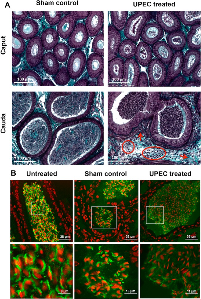
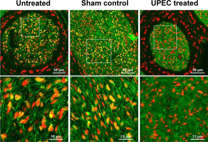
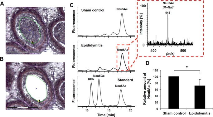
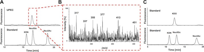
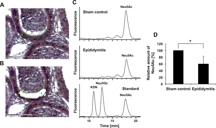
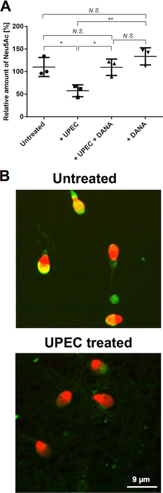
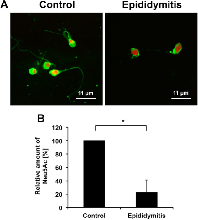
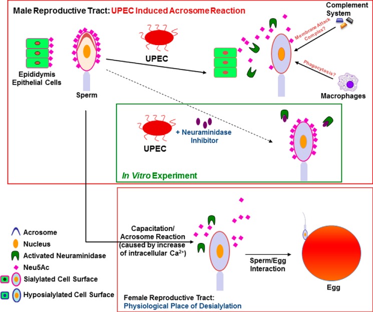
Urinary tract infections caused by uropathogenic Escherichia coli (UPEC) pathovars belong to the most frequent infections in humans. In men, pathogens can also spread to the genital tract via the continuous ductal system, eliciting bacterial prostatitis and/or epididymo-orchitis. Antibiotic treatment usually clears pathogens in acute epididymitis; however, the fertility of patients can be permanently impaired. Because a premature acrosome reaction was observed in an UPEC epididymitis mouse model, and sialidases on the sperm surface are considered to be activated via proteases of the acrosome, we aimed to investigate whether alterations of the sialome of epididymal spermatozoa and surrounding epithelial cells occur during UPEC infection. In UPEC-elicited acute epididymitis in mice, a substantial loss of N-acetylneuraminic acid residues was detected in epididymal spermatozoa and epithelial cells using combined laser microdissection/HPLC-ESI-MS analysis. In support, a substantial reduction of sialic acid residues bound to the surface of spermatozoa was documented in men with a recent history of E. coli-associated epididymitis. In vitro, such an UPEC induced N-acetylneuraminic acid release from human spermatozoa was effectively counteracted by a sialidase inhibitor. These findings strongly suggest a substantial remodeling of the glycocalyx of spermatozoa and epididymal epithelial cells by endogenous sialidases after a premature acrosome reaction during acute epididymitis.
Keywords: bacterial pathogenesis; glycosylation; glycosylation inhibitor; reproduction; sialic acid; sialidase; sperm; spermatozoa.
© 2016 by The American Society for Biochemistry and Molecular Biology, Inc.
Figures
Similar articles
-
Structural and functional integrity of spermatozoa is compromised as a consequence of acute uropathogenic E. coli-associated epididymitis.Biol Reprod. 2013 Sep 19;89(3):59. doi: 10.1095/biolreprod.113.110379. Print 2013 Sep. Biol Reprod. 2013. PMID: 23843239
-
Uropathogenic Escherichia coli causes fibrotic remodelling of the epididymis.J Pathol. 2016 Sep;240(1):15-24. doi: 10.1002/path.4748. J Pathol. 2016. PMID: 27218225
-
Dexamethasone improves therapeutic outcomes in a preclinical bacterial epididymitis mouse model.Hum Reprod. 2019 Jul 8;34(7):1195-1205. doi: 10.1093/humrep/dez073. Hum Reprod. 2019. PMID: 31211847
-
The Immune Characteristics of the Epididymis and the Immune Pathway of the Epididymitis Caused by Different Pathogens.Front Immunol. 2020 Sep 30;11:2115. doi: 10.3389/fimmu.2020.02115. eCollection 2020. Front Immunol. 2020. PMID: 33117332 Free PMC article. Review.
-
Epididymitis: revelations at the convergence of clinical and basic sciences.Asian J Androl. 2015 Sep-Oct;17(5):756-63. doi: 10.4103/1008-682X.155770. Asian J Androl. 2015. PMID: 26112484 Free PMC article. Review.
Cited by
-
Artificial Polysialic Acid Chains as Sialidase-Resistant Molecular-Anchors to Accumulate Particles on Neutrophil Extracellular Traps.Front Immunol. 2017 Sep 29;8:1229. doi: 10.3389/fimmu.2017.01229. eCollection 2017. Front Immunol. 2017. PMID: 29033944 Free PMC article.
-
A Comprehensive Bibliometric Analysis of Orchitis Research from 1980 to 2023.Adv Exp Med Biol. 2025;1469:207-243. doi: 10.1007/978-3-031-82990-1_10. Adv Exp Med Biol. 2025. PMID: 40301259 Review.
-
Epididymal epithelial degeneration and lipid metabolism impairment account for male infertility in occludin knockout mice.Front Endocrinol (Lausanne). 2022 Nov 28;13:1069319. doi: 10.3389/fendo.2022.1069319. eCollection 2022. Front Endocrinol (Lausanne). 2022. PMID: 36518247 Free PMC article.
-
Sialylated Cervical Mucins Inhibit the Activation of Neutrophils to Form Neutrophil Extracellular Traps in Bovine in vitro Model.Front Immunol. 2019 Nov 6;10:2478. doi: 10.3389/fimmu.2019.02478. eCollection 2019. Front Immunol. 2019. PMID: 31781090 Free PMC article.
-
Infectious, inflammatory and 'autoimmune' male factor infertility: how do rodent models inform clinical practice?Hum Reprod Update. 2018 Jul 1;24(4):416-441. doi: 10.1093/humupd/dmy009. Hum Reprod Update. 2018. PMID: 29648649 Free PMC article. Review.
References
-
- Berger R. E., Kessler D., and Holmes K. K. (1987) Etiology and manifestations of epididymitis in young men: correlations with sexual orientation. J. Infect. Dis. 155, 1341–1343 - PubMed
-
- Pilatz A., Hossain H., Kaiser R., Mankertz A., Schüttler C. G., Domann E., Schuppe H. C., Chakraborty T., Weidner W., and Wagenlehner F. (2015) Acute epididymitis revisited: impact of molecular diagnostics on etiology and contemporary guideline recommendations. Eur. Urol. 68, 428–435 - PubMed
-
- Rusz A., Pilatz A., Wagenlehner F., Linn T., Diemer T., Schuppe H. C., Lohmeyer J., Hossain H., and Weidner W. (2012) Influence of urogenital infections and inflammation on semen quality and male fertility. World J. Urol. 30, 23–30 - PubMed
-
- Stammler A., Hau T., Bhushan S., Meinhardt A., Jonigk D., Lippmann T., Pilatz A., Schneider-Hüther I., and Middendorff R. (2015) Epididymitis: ascending infection restricted by segmental boundaries. Hum. Reprod. 30, 1557–1565 - PubMed
-
- Lang T., Dechant M., Sanchez V., Wistuba J., Boiani M., Pilatz A., Stammler A., Middendorff R., Schuler G., Bhushan S., Tchatalbachev S., Wübbeling F., Burger M., Chakraborty T., Mallidis C., and Meinhardt A. (2013) Structural and functional integrity of spermatozoa is compromised as a consequence of acute uropathogenic E. coli-associated epididymitis. Biol. Reprod. 89, 59. - PubMed
Publication types
MeSH terms
Substances
LinkOut - more resources
Full Text Sources
Other Literature Sources
Medical
