. 2016 Jun 24:6:28617.
doi: 10.1038/srep28617.

Myeloid-Specific Blockade of Notch Signaling Attenuates Choroidal Neovascularization through Compromised Macrophage Infiltration and Polarization in Mice

Affiliations

Myeloid-Specific Blockade of Notch Signaling Attenuates Choroidal Neovascularization through Compromised Macrophage Infiltration and Polarization in Mice

Guo-Rui Dou et al. Sci Rep. .

Abstract

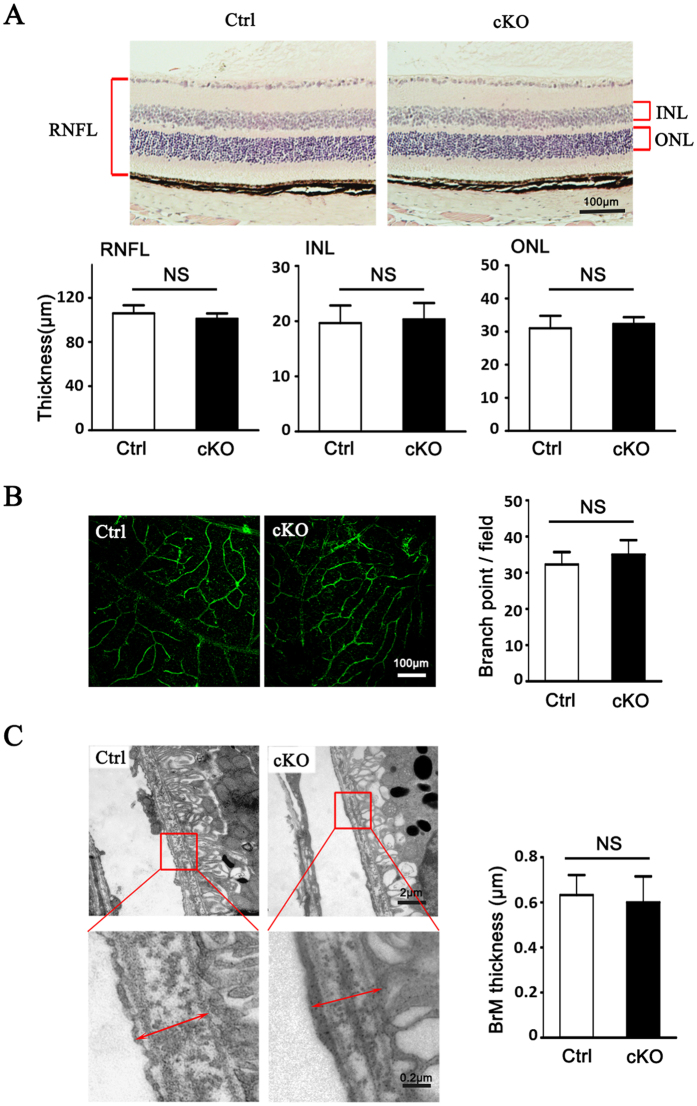
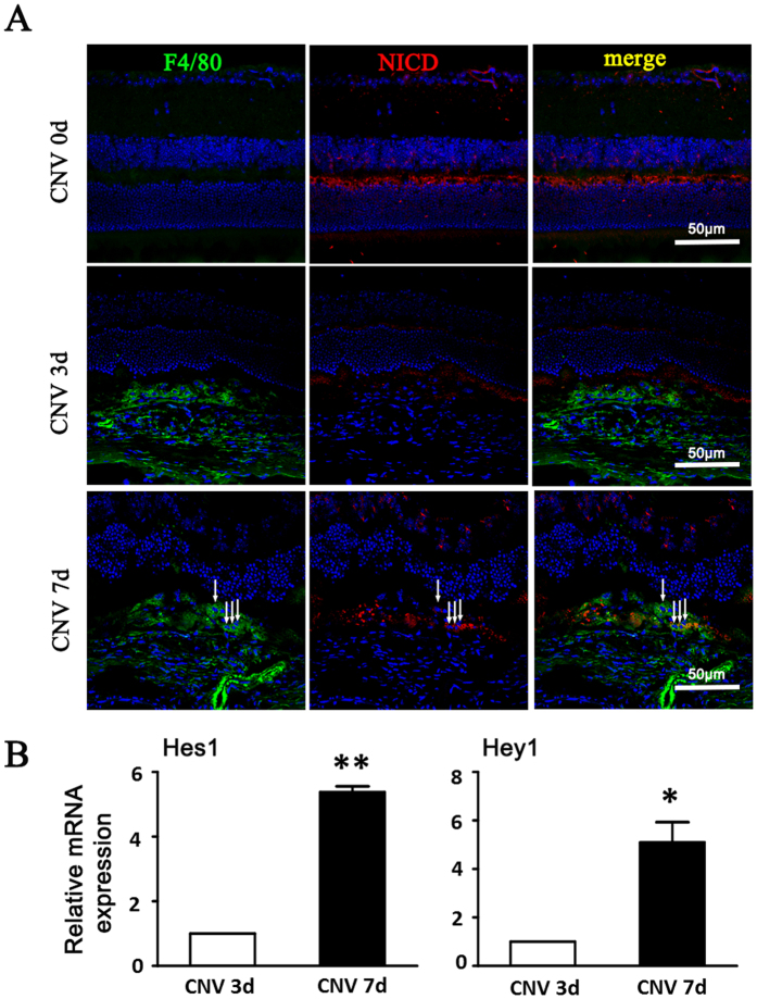
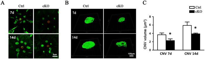
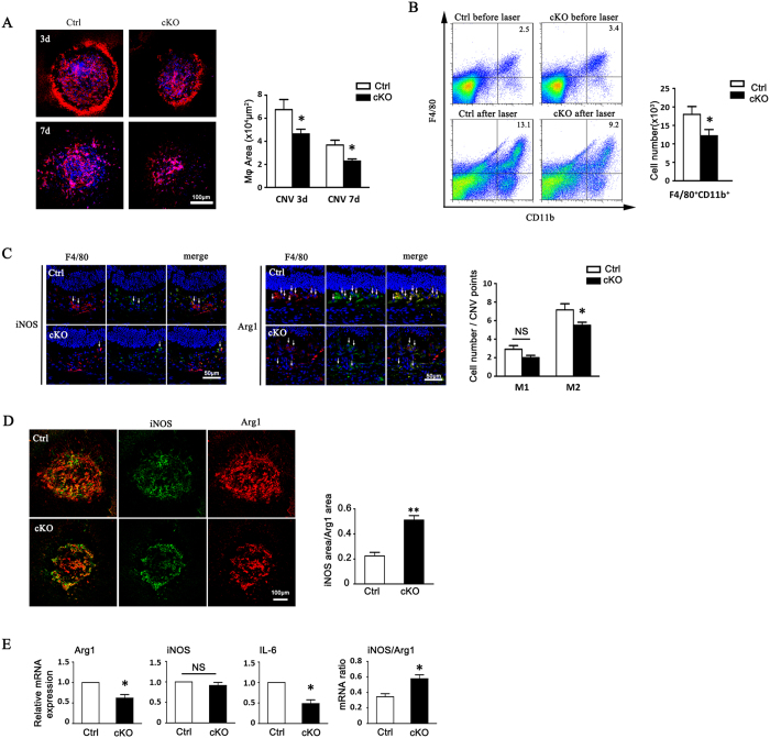
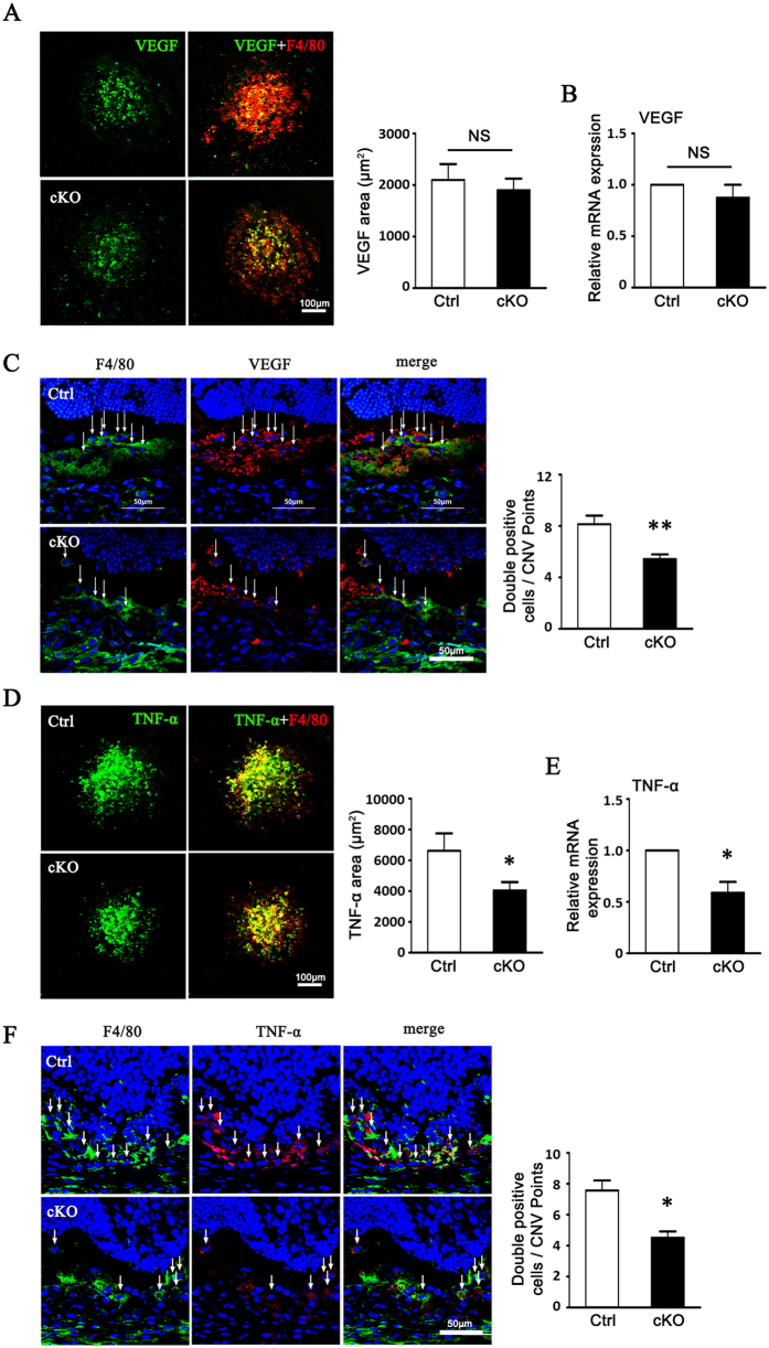
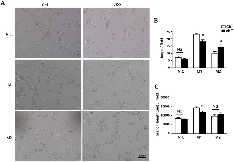
Macrophages have been recognized as an important inflammatory component in choroidal neovascularization (CNV). However, it is unclear how these cells are activated and polarized, how they affect angiogenesis and what the underlining mechanisms are during CNV. Notch signaling has been implicated in macrophage activation. Previously we have shown that inducible disruption of RBP-J, the critical transcription factor of Notch signaling, in adult mice results in enhanced CNV, but it is unclear what is the role of macrophage-specific Notch signaling in the development of CNV. In the current study, by using the myeloid specific RBP-J knockout mouse model combined with the laser-induced CNV model, we show that disruption of Notch signaling in macrophages displayed attenuated CNV growth, reduced macrophage infiltration and activation, and alleviated angiogenic response after laser induction. The inhibition of CNV occurred with reduced expression of VEGF and TNF-α in infiltrating inflammatory macrophages in myeloid specific RBP-J knockout mice. These changes might result in direct inhibition of EC lumen formation, as shown in an in vitro study. Therefore, clinical intervention of Notch signaling in CNV needs to pinpoint myeloid lineage to avoid the counteractive effects of global inhibition.


Figures

Figure 1
Figure 1. Myeloid specific conditional RBP-J knockout (RBP-J cKO) did not result in significant abnormality in eye.
(A) Photomicrographs of the RBP-J cKO (cKO) and control (Ctrl) retina showed similar morphology (H&E). Histograms show the thickness of retinal nerve fiber layer (RNFL), inner nuclear layer (INL), and outer nuclear layer (ONL), respectively. Briefly, three sections of retina tissues with optic nerve were randomly selected from per eye for H&E. The thickness was measured and calculated to get the average thickness, and was compared between two groups. (B) The retinal blood vessels visualized by lectin staining and quantification of the branch points per field. Three representative microscopic fields were examined in each retina and branch points per field were counted. The average number of branch points was calculated. Histogram indicates the comparison the average number of branch points in two groups. (C) Representative transmission electron micrograph of the similar subretinal region of RBP-J cKO and control mice. Briefly, three representative fields were randomly selected from each eye for T.E.M. The average thickness was calculated, and was compared between totally 4 mice per group. Data are presented as mean ± SEM (n = 4 eyes from 4 mice per group). NS, no significance.
Figure 2
Figure 2. Notch activation in the choroidal tissues of mice with induced CNV.
(A) The eyes of wild-type C57/B16 mice were subjected to laser-induced CNV. Eye cups were collected at the indicated time points after laser treatment, and were stained with anti-F4/80 and anti-NICD. Arrows indicated NICD+ macrophages. (B) The two eyes of one mouse were used as one set of RNA retraction. The relative mRNA level of Hes1 and Hey1 was determined by using qRT-PCR, with β-actin as an internal reference control. Each individual experiment was repeated at least three times. Data are presented as mean ± SEM (n = five mice per group). *P < 0.05, **P < 0.01.
Figure 3
Figure 3. RBP-J cKO mice exhibited reduced CNV severity.
RBP-J cKO and control mice were subjected to laser coagulation with six laser burns in one eye (six mice for each group). Choroidal tissues were flat-mounted and stained with FITC-isolectin B4 7 and 14 days later, and examined under a laser-scanning confocal microscope (A), followed by reconstruction of the CNV lesions (B). The CNV volumes were compared between the two groups (C). The optic nerve head are labeled with red circle. Data are presented as mean ± SEM of six RPE/choroid from six mice (six laser burns in each RPE/choroid). *P < 0.05.
Figure 4
Figure 4. Myeloid specific RBP-J deficiency decreased macrophage infiltration and M2 macrophage polarization in CNV lesions.
(A) RBP-J cKO and control mice were subjected to laser coagulation. Choroidal tissues were flat-mounted and stained with anti-F4/80 as indicated time points. Macrophage infiltration area was represented as pixels and the average pixels per CNV lesion were calculated. Histogram shows the comparisons on the average area of macrophage infiltration in five mice per group. (B) Two eyes of one mouse were adopted as one set to prepare the single cell suspensions from RPE-choroidal tissues in (A) at day 3 after laser injury, and analyzed by flow cytometry. The numbers of F4/80+CD11b+ cells were compared between the two groups (five mice per group). (C) Retinal tissues of mice in (A) were immunolabeled with F4/80 and Arg1 or F4/80 and iNOS at day 3 after injury. Arrow indicates Arg1+ macrophages or iNOS+ macrophages. Three representative images per lesion were randomly selected from three biggest CNV lesions in each eye for cell count, and the average number of F4/80+Arg1+ or F4/80+iNOS+ macrophages was calculated and compared in five eyes per group. (D) Flat-mounted choroidal tissues of mice in (C) were immunolabeled with Arg1 and iNOS. The total pixels of iNOS and Arg1 were measured to calculate the ratio of M1/M2. The ratio of M1/M2 was compared between two groups (five eyes per group). (E) Total RNA was prepared from choroidal lysates of RBP-J cKO and control mice at day 3 after laser injury. Two eyes of one mouse were adopted to prepare the RNA retraction for qPCR. The mRNA level of Arg1, iNOS, and IL-6 determined with qRT-PCR, with β-actin as an internal reference control. Each individual experiment was repeated at least three times. The expressions of Arg1, iNOS, IL-6, and the ratio of iNOS/Arg1 were compared. Data are presented as mean ± SEM of five eyes or mice per group. *P < 0.05. NS, no significance.
Figure 5
Figure 5. RBP-J-deficient macrophages exhibited a weakened pro-angiogenic phenotype.
(A) BMDMs were prepared from RBP-J cKO and control mice, and stimulated with PBS (N.C), LPS+IFN-γ (M1) or IL-4 (M2). The expression of iNOS, TNF-α, IL-β, IL-6, VEGF, Arg1, MR and IL-10 mRNA was determined by using qRT-PCR. (B) Culture supernatants of cells in (A) were collected, and the levels of TNF-α, IL-10, IL-1β, IL-6 and VEGF were determined by using ELISA. Each individual experiment was repeated at least three times. Data are presented as mean ± SEM from three mice per group. *P < 0.05, **P < 0.01, ***P < 0.001. NS, no significance.
Figure 6
Figure 6. Myeloid VEGF and TNF-α expressions were reduced in CNV lesions of RBP-J cKO mice.
(A) RBP-J cKO and control mice were subjected to laser coagulation. Choroidal tissues were flat mounted on day 3 and stained with anti-F4/80 and anti-VEGF, and observed under a confocal microscope. The average areas of VEGF expression in 24 randomly selected images were compared between control and RBP-J cKO group. (B) Total RNA of two eyes from one mouse was prepared from choroid tissue in (A). VEGF expression was determined by using qRT-PCR and compared between control and RBP-J cKO group. (C) CNV lesions of mice in (A) were sectioned and stained with anti-F4/80 and anti-VEGF, and observed under a confocal microscope. Five representative images from one eye were randomly selected and F4/80+VEGF+ cells were counted and compared in two groups (five eyes from five mice per group). (D) Flat mounted choroidal tissues in (A) were stained with anti-F4/80 and anti-TNF-α and observed under a confocal microscope. TNF-α immuno-reactivities were compared between control and RBP-J cKO group. (E) Total RNA of two eyes from one mouse was prepared from choroid tissue in (A). TNF-α expression was determined by using qRT-PCR and compared between control and RBP-J cKO group. (F) CNV lesions of mice in (A) were sectioned and stained with anti-F4/80 and anti-TNF-α, and observed under a confocal microscope. F4/80+TNF-α+ cells were compared. Five representative images from one eye were randomly selected and F4/80+ TNF-α+ cells were counted and compared in two groups (five eyes from five mice per group). Data are presented as mean ± SEM (n = five mice per group). *P < 0.05, **P < 0.01. NS, no significance.
Figure 7
Figure 7. Reduced pro-angiogenic phenotype of RBP-J-deficient macrophages inhibited angiogenesis.
(A) BMDMs were prepared from RBP-J cKO and control mice, and stimulated with PBS (N.C), LPS+IFN-γ (M1) or IL-4 (M2). bEND.3 cells were cultured in the presence of conditional medium (CM) from differentially activated macrophages, and induced to form lumens in vitro. Representative images of bEND.3 cells cultured with CM of N.C, M1 or M2 macrophages were shown. (B) Five representative microscopic fields were selected randomly in each culture condition, and the number of lumen loops was calculated and branch length of cell cords of the enclosed lumens was measured. The average number of lumen loops and the average branch length were calculated and compared. Data are presented as mean ± SEM, n = five individual experiments. *P < 0.05. NS, no significance.

Similar articles

Cited by

References

    1. Jager R. D., Mieler W. F. & Miller J. W. Age-related macular degeneration. The New England journal of medicine 358, 2606–2617 (2008). - PubMed
    1. Cherepanoff S., McMenamin P., Gillies M. C., Kettle E. & Sarks S. H. Bruch’s membrane and choroidal macrophages in early and advanced age-related macular degeneration. The British journal of ophthalmology 94, 918–925 (2010). - PubMed
    1. Apte R. S. Regulation of angiogenesis by macrophages. Advances in experimental medicine and biology 664, 15–19 (2010). - PubMed
    1. Skeie J. M. & Mullins R. F. Macrophages in neovascular age-related macular degeneration: friends or foes? Eye (Lond) 23, 747–755 (2009). - PMC - PubMed
    1. Chanmee T., Ontong P., Konno K. & Itano N. Tumor-associated macrophages as major players in the tumor microenvironment. Cancers 6, 1670–1690 (2014). - PMC - PubMed

Publication types

Substances

LinkOut - more resources