. 2016 Aug 1;311(2):G267-75.
doi: 10.1152/ajpgi.00195.2016. Epub 2016 Jun 23.

Hepatocyte nuclear factor 4α is required for cell differentiation and homeostasis in the adult mouse gastric epithelium

Affiliations

Hepatocyte nuclear factor 4α is required for cell differentiation and homeostasis in the adult mouse gastric epithelium

Benjamin D Moore et al. Am J Physiol Gastrointest Liver Physiol. .

Abstract

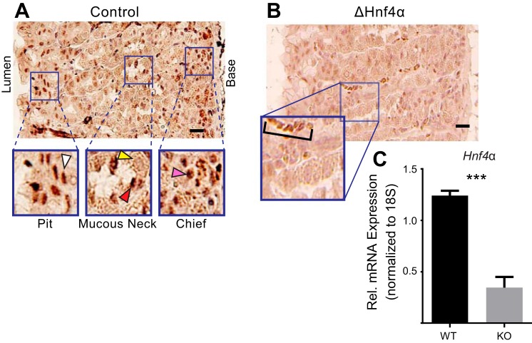
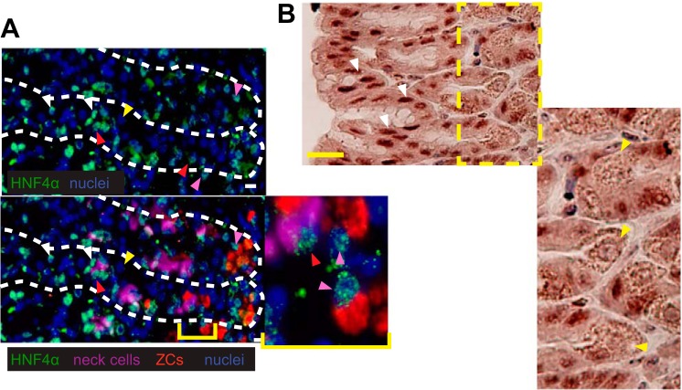
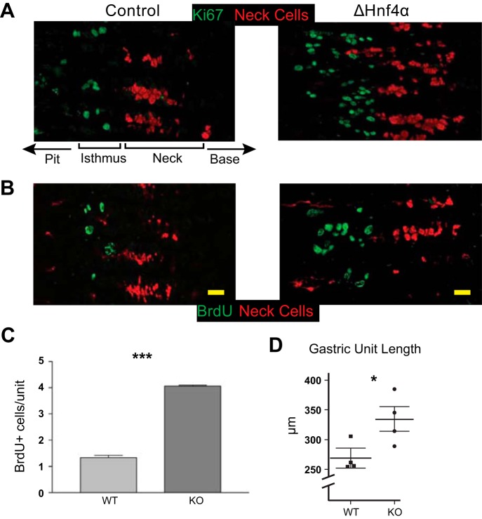
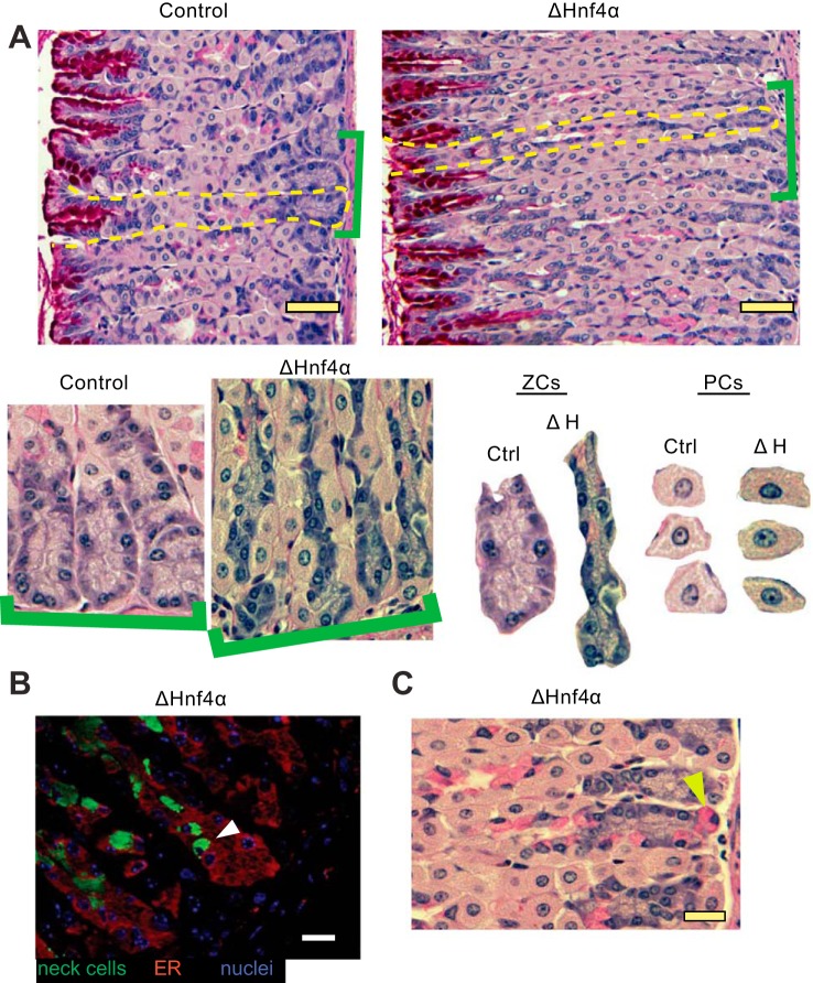
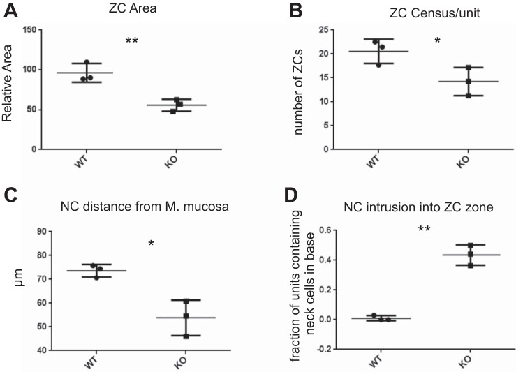
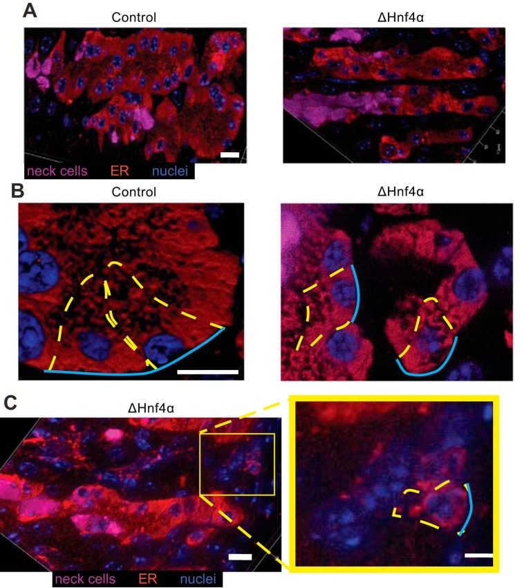
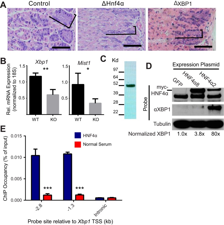
We have previously shown that the sequential transcription factors Xbp1→Mist1 (Bhlha15) govern the ultrastructural maturation of the secretory apparatus in enzyme-secreting zymogenic chief cells (ZCs) in the gastric unit. Here we sought to identify transcriptional regulators upstream of X-box binding protein 1 (XBP1) and MIST1. We used immunohistochemistry to characterize Hnf4α(flox/flox) adult mouse stomachs after tamoxifen-induced deletion of Hnf4α We used qRT-PCR, Western blotting, and chromatin immunoprecipitation to define the molecular interaction between hepatocyte nuclear factor 4 alpha (HNF4α) and Xbp1 in mouse stomach and human gastric cells. We show that HNF4α protein is expressed in pit (foveolar) cells, mucous neck cells, and zymogenic chief cells (ZCs) of the corpus gastric unit. Loss of HNF4α in adult mouse stomach led to reduced ZC size and ER content, phenocopying previously characterized effects of Xbp1 deletion. However, HNF4α(Δ/Δ) stomachs also exhibited additional phenotypes including increased proliferation in the isthmal stem cell zone and altered mucous neck cell migration, indicating a role of HNF4α in progenitor cells as well as in ZCs. HNF4α directly occupies the Xbp1 promoter locus in mouse stomach, and forced HNF4α expression increased abundance of XBP1 mRNA in human gastric cancer cells. Finally, as expected, loss of HNF4α caused decreased Xbp1 and Mist1 expression in mouse stomachs. We show that HNF4α regulates homeostatic proliferation in the gastric epithelium and is both necessary and sufficient for the upstream regulation of the Xbp1→Mist1 axis in maintenance of ZC secretory architecture.

Keywords: mucous neck cell; scaling factor; secretory cells.


Figures

Fig. 1.
Fig. 1.
HNF4α is expressed in the gastric epithelium and can be deleted by Cre recombinase from the adult stomach. A: immunohistochemical stain of HNF4α shows it is expressed in the nuclei of isthmal progenitor and pit cells (white arrowhead), neck cells (red arrowhead), and zymogenic chief cells (pink arrowhead), but not in parietal cells (yellow arrowhead) of the mouse gastric corpus. B: immunohistochemical stain of HNF4α in mice 3 wk postinduction of cre recombinase to delete the floxed Hnf4α allele shows its expression is largely lost vs. control staining in A. Note the background positivity of red blood cells in the inset (bracket) but lack of detectable epithelial cell label (images are representative of n = 4 biological replicates). C: HNF4α mRNA levels in control vs. deleted stomachs. Columns represent means ± SD of three biological replicates; ***P < 0.001. Scale bars = 50 μm.
Fig. 2.
Fig. 2.
Immunofluorescent staining of HNF4α in the gastric epithelium. Images of HNF4α (green) expression in the nucleus (blue) of isthmal progenitor and pit cells (white arrowheads), neck cells (red arrowheads, GSII = purple), and zymogenic cells (pink arrowheads, GIF = red), but again not in parietal cells (yellow arrowheads). Top panel: HNF4α and nuclei; bottom panel: merge with all 4 colors. B: immunohistochemical stain for HNF4α (brown) in region of gastric pit shows strong pit cell nuclear staining (white arrowheads) and only background staining in parietal cells (yellow arrowheads). Scale bars represent 20 μm.
Fig. 3.
Fig. 3.
Loss of HNF4α leads to increased proliferation in the gastric epithelium. A: immunofluorescent staining with Ki67 (green) shows increased proliferation in ΔHNF4α vs. control mouse gastric corpus (mucous neck cells labeled red with GS-II lectin). Note orientation of gastric units with gastric lumen to left and base to right; A and B focus only on isthmus and neck zones. B and C: staining and quantification of BrdU (green) incorporation in control and ΔHNF4α epithelium. Columns = means ± SE, n = 3 biological replicates; ***P < 0.001. D: increased proliferation correlated with longer units. n = 4 mice; mean ± SE; *P < 0.05. Scale bars = 20 μm.
Fig. 4.
Fig. 4.
Loss of HNF4α disrupts the normal differentiation of the mucous neck cell/zymogenic cell lineage. A: H&E + periodic acid Schiff (to detect mucins) of mouse gastric epithelium in control and ΔHNF4α mice. Brackets highlight higher magnification views, below, of bases of gastric units. Isolated bases (labeled “ZCs”) and random PCs from the images are shown side by side at the same magnification. Note that ZCs are smaller in ΔHNF4α mice, whereas PCs do not show morphological changes. Scale bars = 50 μm. B: immunofluorescent stain of the ER marker Calregulin (red) and the mucous neck cell marker GSII (green) in ΔHNF4α mice. Note that mucous neck cells show aberrant localization to the base of the unit, within the ZC zone (white arrowhead), also visible by PAS staining for mucus in C (e.g., yellow arrowhead). Scale bars = 20 μm.
Fig. 5.
Fig. 5.
Quantification of histological data from n = 3 biological replicates. A: ZC size (measured as pixels per cell), normalized to control (WT). B: number of ZCs per unit. C: distance between the mucous neck cell (NC) nearest the base of the gastric unit (i.e., the muscularis mucosa basement membrane) and the base of the unit. D: the mean fraction of units per mouse containing mucous neck cells in the base regions where ZCs reside. *P < 0.05, **P < 0.01 by one-tailed t-test, unequal variance; data expressed as mean from each mouse + range of all 3 mice.
Fig. 6.
Fig. 6.
Zymogenic cell abnormalities in the absence of HNF4α are caused by loss of HNF4α targets regulating ER and the cell cycle. Immunofluorescent stain of the ER marker Calregulin (red) and the mucous neck cell marker GSII (magenta) in control and ΔHNF4α mice (nuclei: blue). Note less elaborate ER network in HNF4α mice (left and right panels at same magnification). A: optical sections. B: higher magnification Z-stack 3-dimensional projection of 20 0.5-μm optical sections in different mice. C: optical section of ΔHNF4α showing occasional bases harboring small ZCs containing markedly scant ER. Representative ZCs outlined in dashed yellow with blue lines showing basement membrane surface. All scale bars = 10 μm.
Fig. 7.
Fig. 7.
HNF4α is required to maintain Xbp1 expression in the mouse stomach. A: H&E staining of mouse gastric epithelium in control, ΔHNF4α and ΔXBP1 mice. As in ΔHNF4α stomachs (Fig. 4A), ZCs are noticeably smaller (brackets outline ZC zone) upon loss of XBP1 (scale bar = 100 μm). B: measurement of mRNA with quantitative-RT-PCR in control and ΔHNF4α mouse gastric corpus shows abundance of Xbp1, and its downstream target Mist1, decrease significantly after deletion of Hnf4α. Means ± SD, n = 3 biological replicates; *P < 0.05, **P < 0.01. C: Western blot of protein isolated from normal mouse stomach and probed with an antibody specific to the P1 splice variant of HNF4α. D: Western blot of AGS gastric cell line transiently transfected with expression plasmids encoding two splice variants of HNF4α or a GFP control shows XBP1 expression is enhanced by overexpression of HNF4α. E: chromatin immunoprecipitation of mouse gastric corpus shows that HNF4α occupies two sites in the Xbp1 promoter compared with immunoprecipitation with normal rabbit serum but not an intronic control region (i.e., with no HNF4α preferred binding sequences predicted). Bars = means ± SD of n = 3 biological replicates, significance was determined via ANOVA with Dunnet's post hoc comparison; ***P < 0.001.

References

    1. Acosta-Alvear D, Zhou Y, Blais A, Tsikitis M, Lents NH, Arias C, Lennon CJ, Kluger Y, Dynlacht BD. XBP1 controls diverse cell type- and condition-specific transcriptional regulatory networks. Mol Cell 27: 53–66, 2007. - PubMed
    1. Ahn SH, Shah YM, Inoue J, Morimura K, Kim I, Yim S, Lambert G, Kurotani R, Nagashima K, Gonzalez FJ, Inoue Y. Hepatocyte nuclear factor 4alpha in the intestinal epithelial cells protects against inflammatory bowel disease. Inflamm Bowel Dis 14: 908–920, 2008. - PMC - PubMed
    1. Anti M, Armuzzi A, Gasbarrini A, Gasbarrini G. Importance of changes in epithelial cell turnover during Helicobacter pylori infection in gastric carcinogenesis. Gut 43, Suppl 1: S27–S32, 1998. - PMC - PubMed
    1. Babeu JP, Darsigny M, Lussier CR, Boudreau F. Hepatocyte nuclear factor 4alpha contributes to an intestinal epithelial phenotype in vitro and plays a partial role in mouse intestinal epithelium differentiation. Am J Physiol Gastrointest Liver Physiol 297: G124–G134, 2009. - PubMed
    1. Battle MA, Konopka G, Parviz F, Gaggl AL, Yang C, Sladek FM, Duncan SA. Hepatocyte nuclear factor 4alpha orchestrates expression of cell adhesion proteins during the epithelial transformation of the developing liver. Proc Natl Acad Sci USA 103: 8419–8424, 2006. - PMC - PubMed

MeSH terms