

Characterization of Pancreatic Cancer Cell Thermal Response to Heat Ablation or Cryoablation
- PMID: 27340260
- PMCID: PMC5616062
- DOI: 10.1177/1533034616655658
Characterization of Pancreatic Cancer Cell Thermal Response to Heat Ablation or Cryoablation
Abstract
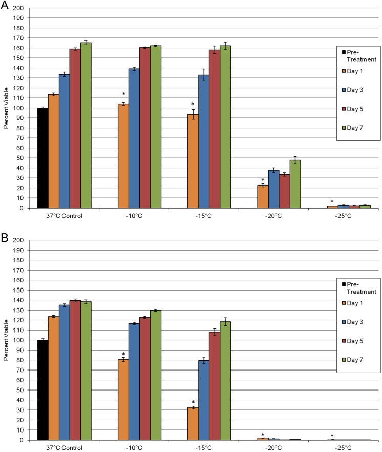
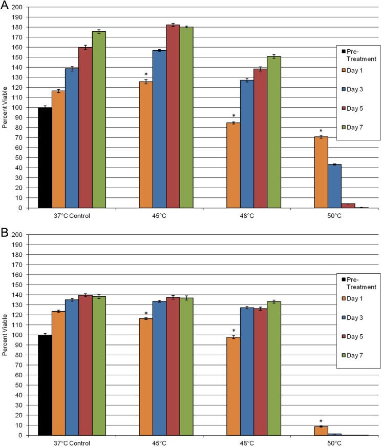
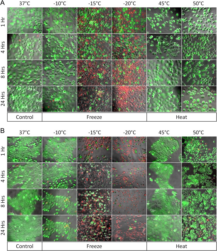
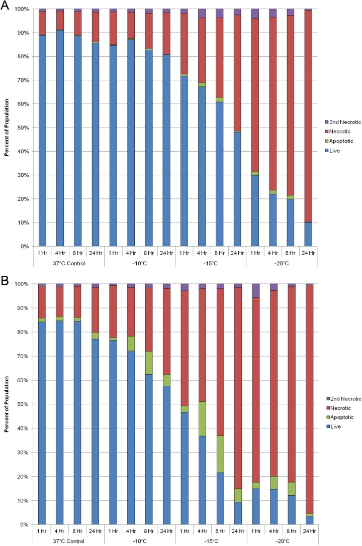
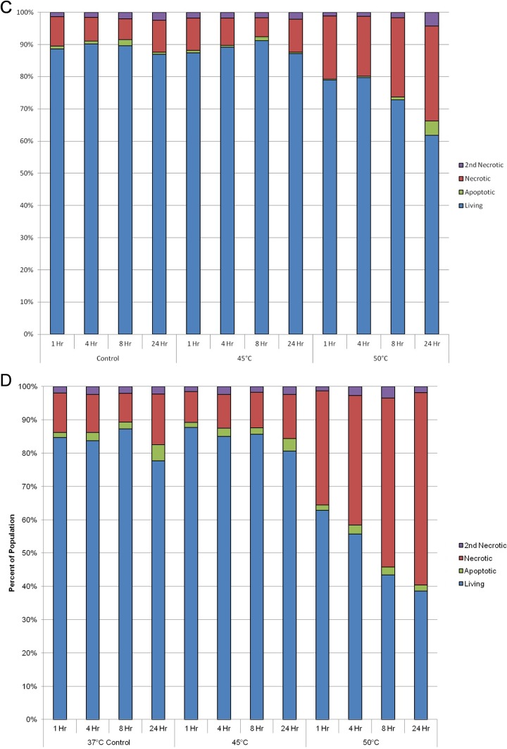
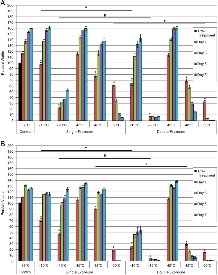
One of the most lethal carcinomas is pancreatic cancer. As standard treatment using chemotherapy and radiation has shown limited success, thermal regimens (cryotherapy or heat ablation) are emerging as viable alternatives. Although promising, our understanding of pancreatic cancer response to thermal ablation remains limited. In this study, we investigated the thermal responses of 2 pancreatic cancer cell lines in an effort to identify the minimum lethal temperature needed for complete cell death to provide guidance for in vivo applications. PANC-1 and BxPC-3 were frozen (-10°C to -25°C) or heated (45°C-50°C) in single and repeated exposure regimes. Posttreatment survival and recovery were analyzed using alamarBlue assay over a 7-day interval. Modes of cell death were assessed using fluorescence microscopy (calcein acetoxymethyl ester/propidium iodide) and flow cytometry (YO-PRO-1/propidium iodide). Freezing to -10°C resulted in minimal cell death. Exposure to -15°C had a mild impact on PANC-1 survival (93%), whereas BxPC-3 was more severely damaged (33%). Exposure to -20°C caused a significant reduction in viability (PANC-1 = 23%; BxPC-3 = 2%) whereas -25°C yielded complete death. Double freezing exposure was more effective than single exposure. Repeat exposure to -15°C resulted in complete death of BxPC-3, whereas -20°C severely impacted PANC-1 (7%). Heating to 45°C resulted in minimum cell death. Exposure to 48°C yielded a slight increase in cell loss (PANC-1 = 85%; BxPC-3 = 98%). Exposure to 50°C caused a significant decline (PANC-1 = 70%; BxPC-3 = 9%) with continued deterioration to 0%. Double heating to 45°C resulted in similar effects observed in single exposures, whereas repeated 48°C resulted in significant increases in cell death (PANC-1 = 68%; BxPC-3 = 29%). In conclusion, we observed that pancreatic cancer cells were completely destroyed at temperatures <-25°C or >50°C using single thermal exposures. Repeated exposures resulted in increased cell death at less extreme temperatures. Our data suggest that thermal ablation strategies (heat or cryoablation) may represent a viable technique for the treatment of pancreatic cancer.
Keywords: apoptosis; cryoablation; hyperthermia; lethal temperature; pancreatic cancer; thermal therapy.
Conflict of interest statement
Figures
Similar articles
-
An In Vitro Investigation into Cryoablation and Adjunctive Cryoablation/Chemotherapy Combination Therapy for the Treatment of Pancreatic Cancer Using the PANC-1 Cell Line.Biomedicines. 2022 Feb 15;10(2):450. doi: 10.3390/biomedicines10020450. Biomedicines. 2022. PMID: 35203660 Free PMC article.
-
Breast Cancer Cryoablation: Assessment of the Impact of Fundamental Procedural Variables in an In Vitro Human Breast Cancer Model.Breast Cancer (Auckl). 2020 Nov 12;14:1178223420972363. doi: 10.1177/1178223420972363. eCollection 2020. Breast Cancer (Auckl). 2020. PMID: 33239880 Free PMC article.
-
In vitro and in vivo anticancer efficacy of silibinin against human pancreatic cancer BxPC-3 and PANC-1 cells.Cancer Lett. 2013 Jun 28;334(1):109-17. doi: 10.1016/j.canlet.2012.09.004. Epub 2012 Sep 26. Cancer Lett. 2013. PMID: 23022268
-
Ablation Strategies for Locally Advanced Pancreatic Cancer.Dig Surg. 2016;33(4):351-9. doi: 10.1159/000445021. Epub 2016 May 25. Dig Surg. 2016. PMID: 27216160 Review.
-
Needle-based ablation of renal parenchyma using microwave, cryoablation, impedance- and temperature-based monopolar and bipolar radiofrequency, and liquid and gel chemoablation: laboratory studies and review of the literature.J Endourol. 2004 Feb;18(1):83-104. doi: 10.1089/089277904322836749. J Endourol. 2004. PMID: 15006061 Review.
Cited by
-
Magnetic Fluid Hyperthermia as Treatment Option for Pancreatic Cancer Cells and Pancreatic Cancer Organoids.Int J Nanomedicine. 2021 Apr 23;16:2965-2981. doi: 10.2147/IJN.S288379. eCollection 2021. Int J Nanomedicine. 2021. PMID: 33935496 Free PMC article.
-
Hyperthermia Enhances Efficacy of Chemotherapeutic Agents in Pancreatic Cancer Cell Lines.Biomolecules. 2022 Apr 29;12(5):651. doi: 10.3390/biom12050651. Biomolecules. 2022. PMID: 35625581 Free PMC article.
-
The Assessment of a Novel Endoscopic Ultrasound-Compatible Cryocatheter to Ablate Pancreatic Cancer.Biomedicines. 2024 Feb 23;12(3):507. doi: 10.3390/biomedicines12030507. Biomedicines. 2024. PMID: 38540120 Free PMC article.
-
Investigating synthetic lethality and PARP inhibitor resistance in pancreatic cancer through enantiomer differential activity.Cell Death Discov. 2025 Mar 16;11(1):106. doi: 10.1038/s41420-025-02382-3. Cell Death Discov. 2025. PMID: 40091075 Free PMC article.
-
In Vitro Measurement and Mathematical Modeling of Thermally-Induced Injury in Pancreatic Cancer Cells.Cancers (Basel). 2023 Jan 21;15(3):655. doi: 10.3390/cancers15030655. Cancers (Basel). 2023. PMID: 36765619 Free PMC article.
References
-
- National Cancer Institute. SEER Cancer Statistics Factsheets: Pancreatic Cancer, 1975-2012. Bethesda, MD: National Cancer Institute; 2015. Website http://seer.cancer.gov/csr/1975_2012/. Published November 2014, Updated April 2015, Accessed October 21, 2015.
-
- National Cancer Institute. PDQ® Pancreatic Cancer Treatment. Bethesda, MD: National Cancer Institute; 2015. Website http://www.cancer.gov/types/pancreatic/patient/pancreatic-treatment-pdq#.... Updated July 2, 2015, Accessed October 21, 2015.
-
- Treating Pancreatic Cancer: How is Pancreatic Cancer Treated? Atlanta, GA: American Cancer Society; Website http://www.cancer.org/cancer/pancreaticcancer/detailedguide/pancreatic-c.... Published June 11, 2014, Updated January 9, 2015, Accessed October 21, 2015.
Publication types
MeSH terms
Grants and funding
LinkOut - more resources
Full Text Sources
Other Literature Sources
Medical
Research Materials
