. 2016:2016:7414036.
doi: 10.1155/2016/7414036. Epub 2016 Jun 2.

Platelet-Rich Plasma Obtained with Different Anticoagulants and Their Effect on Platelet Numbers and Mesenchymal Stromal Cells Behavior In Vitro

Affiliations

Platelet-Rich Plasma Obtained with Different Anticoagulants and Their Effect on Platelet Numbers and Mesenchymal Stromal Cells Behavior In Vitro

Ronaldo José Farias Corrêa do Amaral et al. Stem Cells Int. 2016.

Abstract

There are promising results in the use of platelet-rich plasma (PRP) for musculoskeletal tissue repair. However, the variability in the methodology for its obtaining may cause different and opposing findings in the literature. Particularly, the choice of the anticoagulant is the first definition to be made. In this work, blood was collected with sodium citrate (SC), ethylenediaminetetraacetic acid (EDTA), or anticoagulant citrate dextrose (ACD) solution A, as anticoagulants, prior to PRP obtaining. Hematological analysis and growth factors release quantification were performed, and the effects on mesenchymal stromal cell (MSC) culture, such as cytotoxicity and cell proliferation (evaluated by MTT method) and gene expression, were evaluated. The use of EDTA resulted in higher platelet yield in whole blood; however, it induced an increase in the mean platelet volume (MPV) following the blood centrifugation steps for PRP obtaining. The use of SC and ACD resulted in higher induction of MSC proliferation. On the other hand, PRP obtained in SC presented the higher platelet recovery after the blood first centrifugation step and a minimal change in MSC gene expression. Therefore, we suggest the use of SC as the anticoagulant for PRP obtaining.


Figures

Figure 1
Figure 1
Platelet yield and recovery in blood collected with different anticoagulants. Blood was collected in EDTA, SC, and ACD in five different donors and platelet concentration was quantified (a) as well as platelet recovery after the first centrifugation step (b). An individual analysis between ACD and ACD-2 of platelet recovery was also performed (c). Data are expressed as bar (a), box (b), and dot (c) plots. Similar symbols in (b) correspond to statistic similarity among groups (p > 0.05).
Figure 2
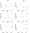
Figure 2
Mean platelet value quantification of samples containing different anticoagulants. Mean platelet value was quantified in five different donors in whole blood (WB), PRP1, and PRP2, in tubes containing EDTA, SC, or ACD solution. The average values of the five different donors are also represented in the figure. “” corresponds to statistical difference between EDTA and SC groups as well as EDTA and ACD groups (p < 0.05).
Figure 3
Figure 3
Growth factor quantification in PRPr obtained in different anticoagulant. TGF-β1 (a) and VEGF quantification (b). Data are expressed as mean, and error bars correspond to standard error.
Figure 4
Figure 4
BM-MSC viability in PRPr obtained with different anticoagulant. Absorbance at 570nm was measured after MTT viability assay of cells cultivated in different PRPr concentrations, obtained with EDTA (a), SC (b), and ACD (c), as well as 10% FBS (control). Data are expressed as mean, and error bars correspond to standard error. “” corresponds to statistical similarity with 10% FBS (p > 0.05).
Figure 5
Figure 5
Photomicrography of BM-MSC. Cells cultivated for eight days in medium supplemented with 10% FBS (a) or a pool from four donors of 5% PRPr obtained from collection tubes containing EDTA (b), SC (c), or ACD (d). Phase contrast, 200x magnification, and scale bars: 50 µm.
Figure 6
Figure 6
Relative gene expression. RUNX2, PPARγ2, SOX9, and Oct-4 gene expression in cells cultured in medium supplemented with 10% FBS (control group) or 5% PRPr obtained with EDTA, SC, or ACD. Data are expressed as relative quantification of gene expression (RQ). Upper and lower error bars correspond to RQ maximum and RQ minimum, respectively.

Similar articles

Cited by

References

    1. Marx R. E. Platelet-rich plasma (PRP): what is PRP and what is not PRP? Implant Dentistry. 2001;10(4):225–228. doi: 10.1097/00008505-200110000-00002. - DOI - PubMed
    1. Filardo G., Kon E., Buda R., et al. Platelet-rich plasma intra-articular knee injections for the treatment of degenerative cartilage lesions and osteoarthritis. Knee Surgery, Sports Traumatology, Arthroscopy. 2011;19(4):528–535. doi: 10.1007/s00167-010-1238-6. - DOI - PubMed
    1. Sánchez M., Azofra J., Anitua E., et al. Plasma rich in growth factors to treat an articular cartilage avulsion: a case report. Medicine and Science in Sports and Exercise. 2003;35(10):1648–1652. doi: 10.1249/01.mss.0000089344.44434.50. - DOI - PubMed
    1. Kon E., Filardo G., Di Matteo B., Marcacci M. PRP for the treatment of cartilage pathology. The Open Orthopaedics Journal. 2013;7:120–128. - PMC - PubMed
    1. Taylor D. W., Petrera M., Hendry M., Theodoropoulos J. S. A systematic review of the use of platelet-rich plasma in sports medicine as a new treatment for tendon and ligament injuries. Clinical Journal of Sport Medicine. 2011;21(4):344–352. doi: 10.1097/jsm.0b013e31821d0f65. - DOI - PubMed