

Amla Enhances Mitochondrial Spare Respiratory Capacity by Increasing Mitochondrial Biogenesis and Antioxidant Systems in a Murine Skeletal Muscle Cell Line
- PMID: 27340504
- PMCID: PMC4909908
- DOI: 10.1155/2016/1735841
Amla Enhances Mitochondrial Spare Respiratory Capacity by Increasing Mitochondrial Biogenesis and Antioxidant Systems in a Murine Skeletal Muscle Cell Line
Abstract
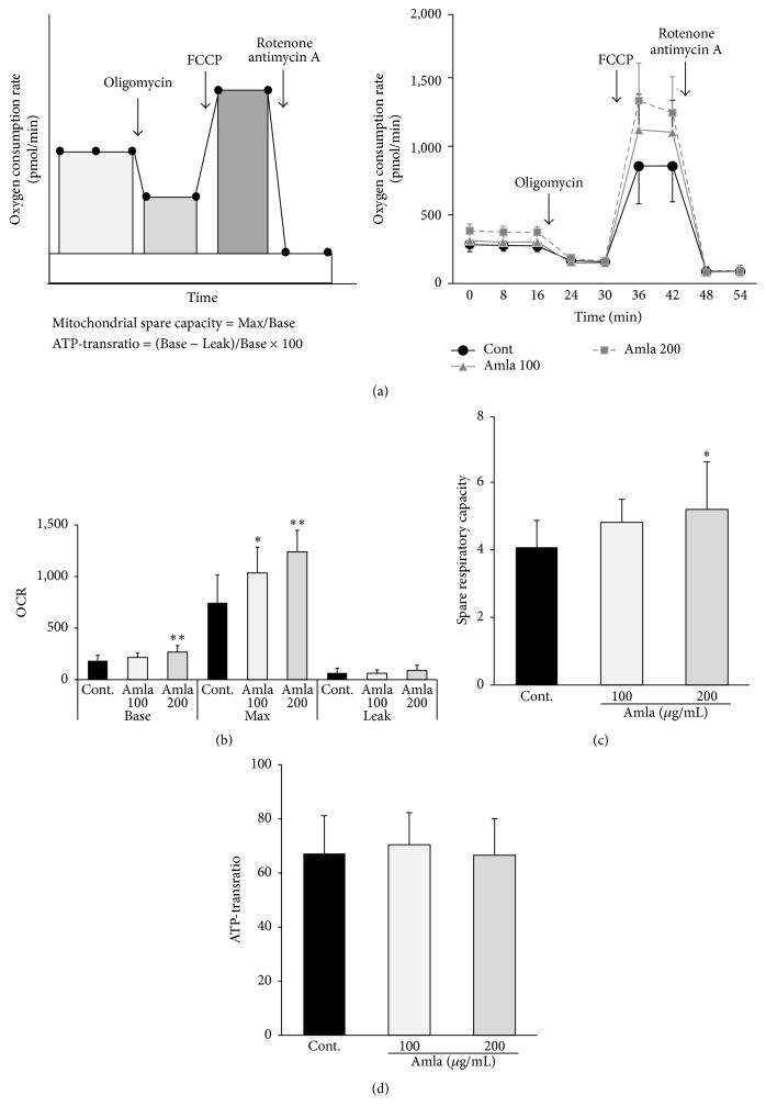
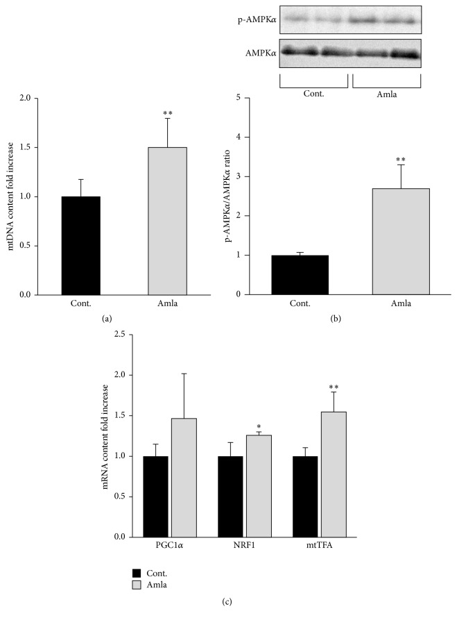
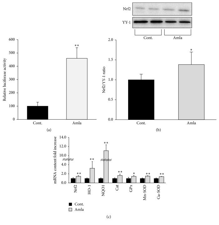
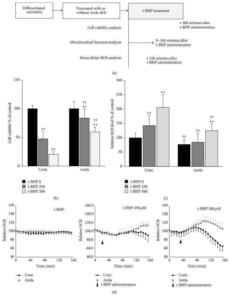
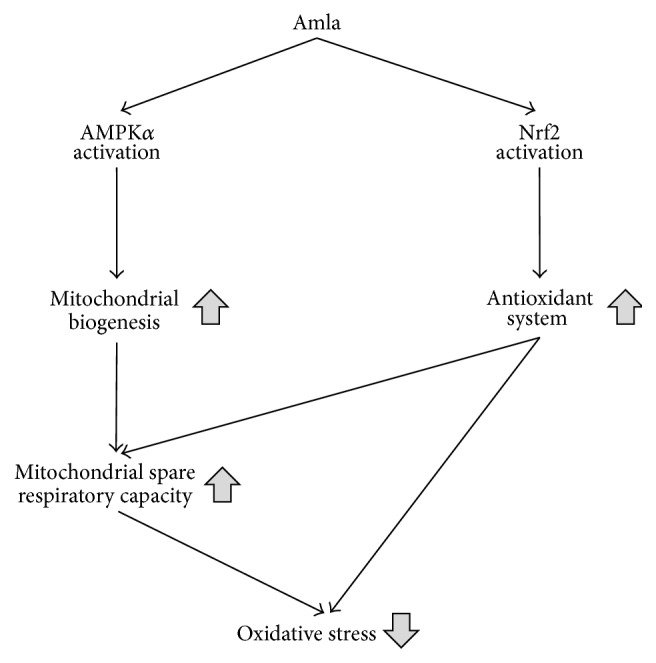
Amla is one of the most important plants in Indian traditional medicine and has been shown to improve various age-related disorders while decreasing oxidative stress. Mitochondrial dysfunction is a proposed cause of aging through elevated oxidative stress. In this study, we investigated the effects of Amla on mitochondrial function in C2C12 myotubes, a murine skeletal muscle cell model with abundant mitochondria. Based on cell flux analysis, treatment with an extract of Amla fruit enhanced mitochondrial spare respiratory capacity, which enables cells to overcome various stresses. To further explore the mechanisms underlying these effects on mitochondrial function, we analyzed mitochondrial biogenesis and antioxidant systems, both proposed regulators of mitochondrial spare respiratory capacity. We found that Amla treatment stimulated both systems accompanied by AMPK and Nrf2 activation. Furthermore, we found that Amla treatment exhibited cytoprotective effects and lowered reactive oxygen species (ROS) levels in cells subjected to t-BHP-induced oxidative stress. These effects were accompanied by increased oxygen consumption, suggesting that Amla protected cells against oxidative stress by using enhanced spare respiratory capacity to produce more energy. Thus we identified protective effects of Amla, involving activation of mitochondrial function, which potentially explain its various effects on age-related disorders.
Figures
Similar articles
-
Metabolomic Analysis and Antioxidant Effect of Amla (Emblica officinalis) Extract in Preventing Oxidative Stress-Induced Red Cell Damage and Plasma Protein Alterations: An In Vitro Study.J Med Food. 2018 Jan;21(1):81-89. doi: 10.1089/jmf.2017.3942. Epub 2017 Oct 24. J Med Food. 2018. PMID: 29064307
-
Antiproliferative effect of silver nanoparticles synthesized using amla on Hep2 cell line.Asian Pac J Trop Med. 2013 Jan;6(1):1-10. doi: 10.1016/S1995-7645(12)60193-X. Asian Pac J Trop Med. 2013. PMID: 23317879
-
Amla (Emblica officinalis Gaertn.) attenuates age-related renal dysfunction by oxidative stress.J Agric Food Chem. 2007 Sep 19;55(19):7744-52. doi: 10.1021/jf072105s. Epub 2007 Aug 23. J Agric Food Chem. 2007. PMID: 17715896
-
The role of mitochondria in aging of skeletal muscle.Biogerontology. 2008 Apr;9(2):67-84. doi: 10.1007/s10522-007-9121-7. Epub 2008 Jan 4. Biogerontology. 2008. PMID: 18175203 Review.
-
Hepatoprotective properties of the Indian gooseberry (Emblica officinalis Gaertn): a review.Food Funct. 2013 Oct;4(10):1431-41. doi: 10.1039/c3fo60237k. Food Funct. 2013. PMID: 23978895 Review.
Cited by
-
Ultra-Small Iron Nanoparticles Target Mitochondria Inducing Autophagy, Acting on Mitochondrial DNA and Reducing Respiration.Pharmaceutics. 2021 Jan 12;13(1):90. doi: 10.3390/pharmaceutics13010090. Pharmaceutics. 2021. PMID: 33445442 Free PMC article.
-
Suppression of protein degradation by leucine requires its conversion to β-hydroxy-β-methyl butyrate in C2C12 myotubes.Aging (Albany NY). 2019 Dec 24;11(24):11922-11936. doi: 10.18632/aging.102509. Epub 2019 Dec 24. Aging (Albany NY). 2019. PMID: 31881014 Free PMC article.
-
Using Human 'Personalized' Cybrids to Identify Drugs/Agents That Can Regulate Chronic Lymphoblastic Leukemia Mitochondrial Dysfunction.Int J Mol Sci. 2023 Jul 3;24(13):11025. doi: 10.3390/ijms241311025. Int J Mol Sci. 2023. PMID: 37446202 Free PMC article.
-
Evaluating the Bioenergetics Health Index Ratio in Leigh Syndrome Fibroblasts to Understand Disease Severity.Int J Mol Sci. 2021 Sep 26;22(19):10344. doi: 10.3390/ijms221910344. Int J Mol Sci. 2021. PMID: 34638685 Free PMC article.
-
Comparative analysis of the mitochondrial morphology, energy metabolism, and gene expression signatures in three types of blastocyst-derived stem cells.Redox Biol. 2020 Feb;30:101437. doi: 10.1016/j.redox.2020.101437. Epub 2020 Jan 20. Redox Biol. 2020. PMID: 31981893 Free PMC article.
References
-
- Patel S. S., Goyal R. K. Emblica officinalis Geart.: a comprehensive review on phytochemistry, pharmacology and ethnomedicinal uses. Research Journal of Medicinal Plant. 2012;6(1):6–16. doi: 10.3923/rjmp.2012.6.16. - DOI
-
- Bhandari P., Kamdod M. Emblica officinalis (Amla): a review of potential therapeutic applications. International Journal of Green Pharmacy. 2012;6(4):257–269. doi: 10.4103/0973-8258.108204. - DOI
-
- Usharani P., Fatima N., Muralidhar N. Effects of Phyllanthus emblica extract on endothelial dysfunction and biomarkers of oxidative stress in patients with type 2 diabetes mellitus: a randomized, double-blind, controlled study. Diabetes, Metabolic Syndrome and Obesity: Targets and Therapy. 2013;6:275–284. doi: 10.2147/dmso.s46341. - DOI - PMC - PubMed
-
- Bhattacharya A., Chatterjee A., Ghosal S., Bhattacharya S. K. Antioxidant activity of active tannoid principles of Emblica officinalis (amla) Indian Journal of Experimental Biology. 1999;37(7):676–680. - PubMed
-
- Damodara Reddy V., Padmavathi P., Gopi S., Paramahamsa M., Varadacharyulu N. C. Protective effect of Emblica officinalis against alcohol-induced hepatic injury by ameliorating oxidative stress in rats. Indian Journal of Clinical Biochemistry. 2010;25(4):419–424. doi: 10.1007/s12291-010-0058-2. - DOI - PMC - PubMed
MeSH terms
Substances
LinkOut - more resources
Full Text Sources
Other Literature Sources
Medical
