Review
. 2016:2016:6701324.
doi: 10.1155/2016/6701324. Epub 2016 Jun 2.

Intracellular Calcium Dysregulation: Implications for Alzheimer's Disease

Affiliations
Review

Intracellular Calcium Dysregulation: Implications for Alzheimer's Disease

Simona Magi et al. Biomed Res Int. 2016.

Abstract

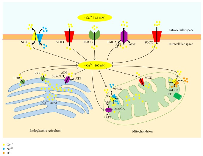
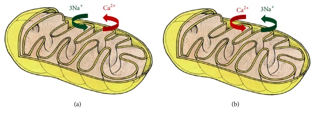
Alzheimer's Disease (AD) is a neurodegenerative disorder characterized by progressive neuronal loss. AD is associated with aberrant processing of the amyloid precursor protein, which leads to the deposition of amyloid-β plaques within the brain. Together with plaques deposition, the hyperphosphorylation of the microtubules associated protein tau and the formation of intraneuronal neurofibrillary tangles are a typical neuropathological feature in AD brains. Cellular dysfunctions involving specific subcellular compartments, such as mitochondria and endoplasmic reticulum (ER), are emerging as crucial players in the pathogenesis of AD, as well as increased oxidative stress and dysregulation of calcium homeostasis. Specifically, dysregulation of intracellular calcium homeostasis has been suggested as a common proximal cause of neural dysfunction in AD. Aberrant calcium signaling has been considered a phenomenon mainly related to the dysfunction of intracellular calcium stores, which can occur in both neuronal and nonneuronal cells. This review reports the most recent findings on cellular mechanisms involved in the pathogenesis of AD, with main focus on the control of calcium homeostasis at both cytosolic and mitochondrial level.


Figures

Figure 1
Figure 1
Intracellular calcium homeostasis. Intracellular calcium levels are tightly regulated within a narrow physiological range [23]. Cellular calcium influx through the plasma membrane is largely mediated by receptor-operated calcium channels (ROCC), voltage-operated calcium channels (VOCC), store-operated calcium channels (SOCC) and, under exceptional circumstances, the sodium/calcium exchanger (NCX). Under physiological conditions, NCX is mainly involved in calcium efflux; however it can also reverse its mode of operation (reverse mode exchange) thereby contributing to calcium influx, especially during strong depolarization and in the presence of high intracellular sodium concentrations [24]. Calcium may also be released into the cytoplasm from the endoplasmic reticulum, through inositol-1,4,5-trisphosphate (IP3R) and ryanodine receptors (RYR). Different systems operate within the cell to counterbalance the cytosolic calcium increase. Specifically, the plasma membrane calcium pump (PMCA), NCX, and sarcoendoplasmic reticulum calcium ATPase (SERCA) participate in restoring physiological calcium levels. The excess of intracellular calcium can also be taken up by mitochondria through the mitochondrial calcium uniporter (MCU). Calcium can be released back into the cytosol through the activity of mitochondrial NCX (mNCX), which can also reverse its mode of operation allowing the access of calcium ions into the mitochondrial matrix. Recently, the mitochondrial hydrogen/calcium exchanger (mHCX) has been proposed to be an electrogenic 1 : 1 mitochondrial calcium/hydrogen antiporter that drives the uptake of calcium into mitochondria at nanomolar cytosolic calcium concentrations [25]. PTP, permeability transition pore; MMCA, mitochondrial membrane Ca2+ATPase.
Figure 2
Figure 2
Modes of operation of mNCX. The figure reports the prevalent modes of operation of mNCX. (a) shows the forward mode of operation of the exchanger, which is prevalent in physiological conditions. In this mode of operation, mNCX mediates the extrusion of calcium ions from mitochondrial matrix in exchange for sodium ions. (b) shows mNCX reverse mode of operation. In this mode of operation, the mitochondrial exchanger mediates the influx of calcium ions into the matrix and the extrusion of sodium ions. The figure has been entirely reproduced from Castaldo et al., 2009 [26], upon written authorization by the editor.
Figure 3
Figure 3
NCXs labeling patterns in neuronal mitochondria ((a)–(i)). (a and d) NCX1-ir mitochondria (arrows) in distal dendrites (CA1 stratum radiatum). ((b) and (e)) NCX2-ir mitochondria (arrows) in hippocampal (b) and neocortical (e) dendrites. (c) NCX3-ir mitochondria in neocortical distal dendrite (arrow). (f) NCX2-positive mitochondrion in a CA1 axon terminal. ((g) and (h)) NCX2 and NCX3-ir mitochondria (arrows) in a cell body (from CA1 pyramidal cell layer); enlarged in the inset in (h), two labeled organelles (arrows) near nuclear envelope. (i) NCX3-ir in neocortical distal dendrite with unlabeled mitochondria (open arrow). In (b) and (d) dendrites are contacted by axon terminals forming asymmetric junction (triangles). In (a) and (e), note the labeling bridging plasma membrane and mitochondrial profile. Open arrows indicate unlabeled mitochondria in dendrites ((a) and (i)) and axon terminals ((b) and (d)). With asterisks the postsynaptic specializations are indicated and the arrowheads show the labeling between mitochondria and plasma membrane. axt, axon terminal; den, dendrite; nu, nucleus; cyt, cytoplasm; sp, dendritic spine. Immunoperoxidase reaction in (a)–(c), (f), (g), and (h) and silver-enhanced immunogold in (d), (e), and (i). Calibration bars: in (a), 0.25 m for (a), (b), (d), (f), and (i); in (a), 0.5 m for inset in (h); in (c), 0.25 m for (c) and (e); in (c), 0.5 m for (g); in (c), 1 m for (h). The figure has been entirely reproduced from Gobbi et al., 2007 [27], upon written authorization by the editor.
Figure 4
Figure 4
NCXs labeling patterns in astrocytic mitochondria ((a)–(d)). (a) NCX3-expressing mitochondrion (arrow) in neocortical astrocytic process; an adjacent dendrite contains two labeled mitochondria (arrows). (b) NCX2-ir mitochondrion (arrow) in hippocampal glial process. Intense labeling is present on plasma membrane. (c) NCX3-labelled sub-plasma membrane mitochondria (arrows) in two astrocytic processes contacting synaptic structures; labeling between a mitochondrion and the plasma membrane is evident (arrowhead). An unlabeled mitochondrion is localized in a dentritic structure (open arrow). (d) A NCX1-unlabeled mitochondrion (open arrow) in a labeled distal astrocytic process in neocortex. Note some positive distal dendrites with unlabeled mitochondria. Open arrows indicate unlabeled mitochondria; triangles show the mitochondrial labeling near the synaptic membrane. Asp, astrocytic process; den, dendrite; axt, axon terminal; sp, spine apparatus. Immunoperoxidase reaction in (b) and (c) and silver-enhanced immunogold in (a). Calibration bars: in (a), 0.25 m for (a), (b), and (c); in (a), 0.5 m for (d). The figure has been entirely reproduced from Gobbi et al., 2007 [27], upon written authorization by the editor.

References

    1. Ballard C., Gauthier S., Corbett A., Brayne C., Aarsland D., Jones E. Alzheimer's disease. The Lancet. 2011;377(9770):1019–1031. doi: 10.1016/s0140-6736(10)61349-9. - DOI - PubMed
    1. Reitz C., Mayeux R. Alzheimer disease: epidemiology, diagnostic criteria, risk factors and biomarkers. Biochemical Pharmacology. 2014;88(4):640–651. doi: 10.1016/j.bcp.2013.12.024. - DOI - PMC - PubMed
    1. Alzheimer's Association. 2013 Alzheimer's disease facts and figures. Alzheimer's & Dementia. 2013;9(2):208–245. doi: 10.1016/j.jalz.2013.02.003. - DOI - PubMed
    1. Bonomo S. M., Rigamonti A. E., Giunta M., et al. Menopausal transition: a possible risk factor for brain pathologic events. Neurobiology of Aging. 2009;30(1):71–80. doi: 10.1016/j.neurobiolaging.2007.05.017. - DOI - PubMed
    1. Pike C. J., Carroll J. C., Rosario E. R., Barron A. M. Protective actions of sex steroid hormones in Alzheimer's disease. Frontiers in Neuroendocrinology. 2009;30(2):239–258. doi: 10.1016/j.yfrne.2009.04.015. - DOI - PMC - PubMed

MeSH terms

LinkOut - more resources