. 2016 Jun 13;3(4):e251.
doi: 10.1212/NXI.0000000000000251. eCollection 2016 Aug.

Next-generation sequencing in neuropathologic diagnosis of infections of the nervous system

Affiliations

Next-generation sequencing in neuropathologic diagnosis of infections of the nervous system

Steven L Salzberg et al. Neurol Neuroimmunol Neuroinflamm. .

Abstract

Objective: To determine the feasibility of next-generation sequencing (NGS) microbiome approaches in the diagnosis of infectious disorders in brain or spinal cord biopsies in patients with suspected CNS infections.

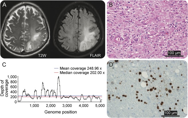
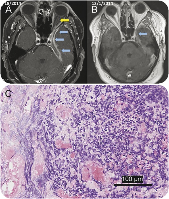
Methods: In a prospective pilot study, we applied NGS in combination with a new computational analysis pipeline to detect the presence of pathogenic microbes in brain or spinal cord biopsies from 10 patients with neurologic problems indicating possible infection but for whom conventional clinical and microbiology studies yielded negative or inconclusive results.

Results: Direct DNA and RNA sequencing of brain tissue biopsies generated 8.3 million to 29.1 million sequence reads per sample, which successfully identified with high confidence the infectious agent in 3 patients for whom validation techniques confirmed the pathogens identified by NGS. Although NGS was unable to identify with precision infectious agents in the remaining cases, it contributed to the understanding of neuropathologic processes in 5 others, demonstrating the power of large-scale unbiased sequencing as a novel diagnostic tool. Clinical outcomes were consistent with the findings yielded by NGS on the presence or absence of an infectious pathogenic process in 8 of 10 cases, and were noncontributory in the remaining 2.

Conclusions: NGS-guided metagenomic studies of brain, spinal cord, or meningeal biopsies offer the possibility for dramatic improvements in our ability to detect (or rule out) a wide range of CNS pathogens, with potential benefits in speed, sensitivity, and cost. NGS-based microbiome approaches present a major new opportunity to investigate the potential role of infectious pathogens in the pathogenesis of neuroinflammatory disorders.


Figures

Figure 1.
Figure 1.. Heatmap shows the top microbial species in each of the 11 samples
All microbial species that explain 10% or more of microbial reads (excluding contaminants) in any sample are shown. Shaded squares indicate the relative proportion of reads from each species in that sample, ranging from blue (1%) to red (100%). Green boxes across the bottom indicate the 3 samples for which the sequence-based diagnosis was independently confirmed.
Figure 2.
Figure 2.. A patient with multifocal nodular lesions diagnosed with CNS tuberculosis
(A) Prebiopsy brain MRI in patient PT-8 shows multifocal subcortical and deep white matter lesions in fluid-attenuated inversion recovery (FLAIR) MRI. Several focal nodular enhancing lesions are seen in T1-weighted + gadolinium (Gad) MRI. A brain biopsy targeted a nodular lesion in the left posterior frontal-parietal region (yellow circles). Subsequent brain MRIs demonstrate a dramatic improvement of the lesions 23 days after biopsy and 15 days after instauration of the anti-TB medications. (B) Diagrammatic representation of the time profile for biopsy processing for neuropathology and next-generation sequencing (NGS) diagnosis in PD-8. Although histologic studies were able to establish the presence of a granulomatous inflammation immediately after biopsy, a more comprehensive neurohistologic analysis completed 6 days after biopsy failed to demonstrate infectious pathogens. NGS diagnosis was established 7 days after biopsy, confirming the presence of Mycobacterium tuberculosis, a finding that was used to support the treatment with antituberculous medication and ruled out the presence of Nocardia infection. (C) Histologic demonstration of granulomatous inflammation in hematoxylin & eosin stains. (D) Grocott methenamine silver stain is negative for fungal species. Similarly, Ziehl-Neelsen stain for acid-fast bacilli (E) is negative.
Figure 3.
Figure 3.. Brain imaging and neuropathologic studies in patient PT-5 diagnosed with progressive multifocal leukoencephalopathy
(A) MRI in patient PT-5 shows an extensive area of signal abnormalities in both T2-weighted and fluid-attenuated inversion recovery (FLAIR) MRI sequences that involved the white matter of the left posterior region of the frontal-parietal lobes. (B) Histologic changes observed in hematoxylin & eosin stain demonstrated extensive gliosis, presence of gemystocytic astrocytes, and mild inflammatory reaction. (C) Next-generation sequencing (NGS) analysis reveals 8,944 JC polyomavirus reads out of 8,954 reads from all viruses. The whole genome of JC polyomavirus was covered by the reads, at an average depth over 200. The NGS findings are consistent with a diagnosis of progressive multifocal leukoencephalopathy (PML). (D) Immunostaining with SV-40 antibodies, a surrogate for JC virus identification, shows several immunopositive nuclei as confirmation of JC virus infection. These findings confirm the diagnosis of PML.
Figure 4.
Figure 4.. Brain imaging and neuropathologic demonstration of Epstein-Barr virus (EBV) encephalitis in patient PT-10
(A–C) Brain MRI shows a focal area of cortical and subcortical signal intensity abnormality. T1-weighted sequences enhanced with gadolinium show a ring-enhanced lesion with perilesional edema, which was the target for the brain biopsy. (D) Histologic demonstration of a granulomatous and lymphohistocytic inflammation with foci of necrosis (hematoxylin & eosin stain) in patient PT-10. (E) In situ hybridization for EBV-encoded RNA in the brain biopsy tissues, which validates the presence of EBV as it had been established by next-generation sequencing and confirms the diagnosis of EBV encephalitis. FLAIR = fluid-attenuated inversion recovery.
Figure 5.
Figure 5.. A patient with focal pachymeningitis and Tolosa-Hunt-like syndrome
(A) Neuroimaging studies in patient PT-2 demonstrate the presence of pachymeningeal and leptomeningeal enhancement (blue arrows) localized in the medial aspect of the left middle cranial fossae extending to the orbital apex with involvement of the dural margin of the left cavernous sinus, Meckel cave, and foramen ovale. Enhancement of the left optic nerve dura (yellow arrow) is also noted in the March 18, 2014, MRI. (B) Neuroimaging studies following 5 weeks of antibiotic treatment (December 1, 2014) show decreased meningeal and dural enhancement along the anteromedial left temporal lobe margin and tentorial leaflet as well as optic nerve dural sheath. (C) Histopathologic studies of the dura and adjacent bone show chronic inflammation comprising lymphocytes, macrophages, and a few plasma cells (hematoxylin & eosin stain).

References

    1. Glaser CA, Honarmand S, Anderson LJ, et al. Beyond viruses: clinical profiles and etiologies associated with encephalitis. Clin Infect Dis 2006;43:1565–1577. - PubMed
    1. Barzon L, Lavezzo E, Militello V, Toppo S, Palu G. Applications of next-generation sequencing technologies to diagnostic virology. Int J Mol Sci 2011;12:7861–7884. - PMC - PubMed
    1. Yin L, Liu L, Sun Y, et al. High-resolution deep sequencing reveals biodiversity, population structure, and persistence of HIV-1 quasispecies within host ecosystems. Retrovirology 2012;9:108. - PMC - PubMed
    1. Lecuit M, Eloit M. The human virome: new tools and concepts. Trends Microbiol 2013;21:510–515. - PMC - PubMed
    1. Wylie KM, Weinstock GM, Storch GA. Virome genomics: a tool for defining the human virome. Curr Opinion Microbiol 2013;16:479–484. - PMC - PubMed

LinkOut - more resources