. 2016 Nov;34(11):2744-2757.
doi: 10.1002/stem.2442. Epub 2016 Jul 17.

Local CXCR4 Upregulation in the Injured Arterial Wall Contributes to Intimal Hyperplasia

Affiliations

Local CXCR4 Upregulation in the Injured Arterial Wall Contributes to Intimal Hyperplasia

Xudong Shi et al. Stem Cells. 2016 Nov.

Abstract

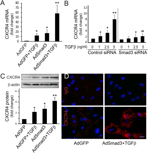
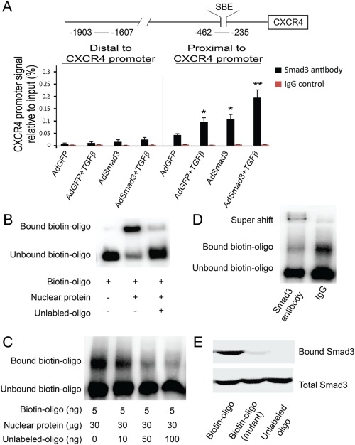
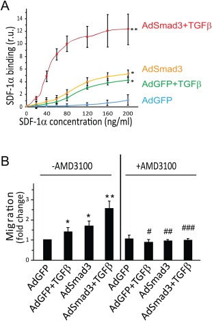
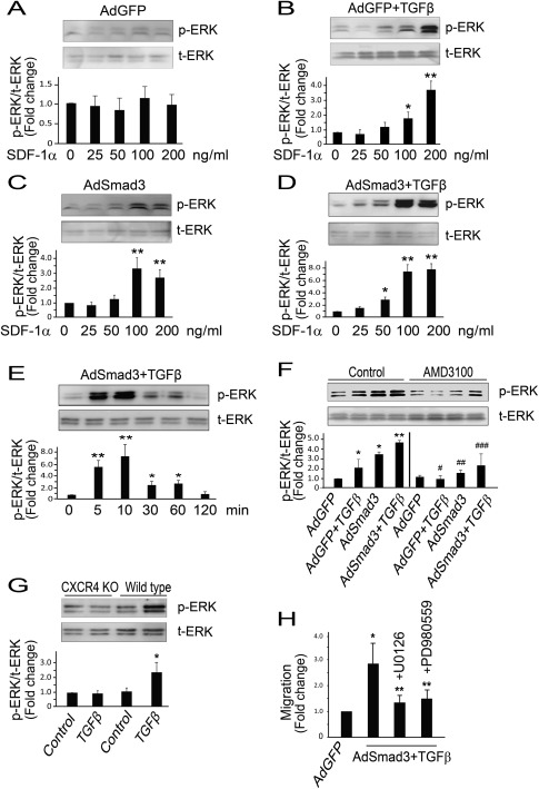
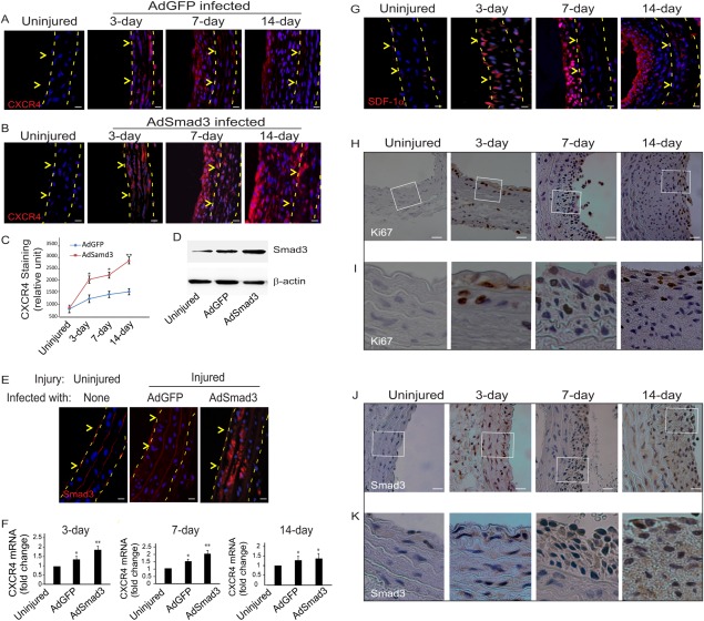
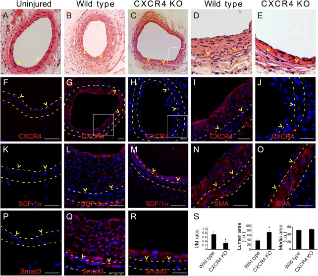
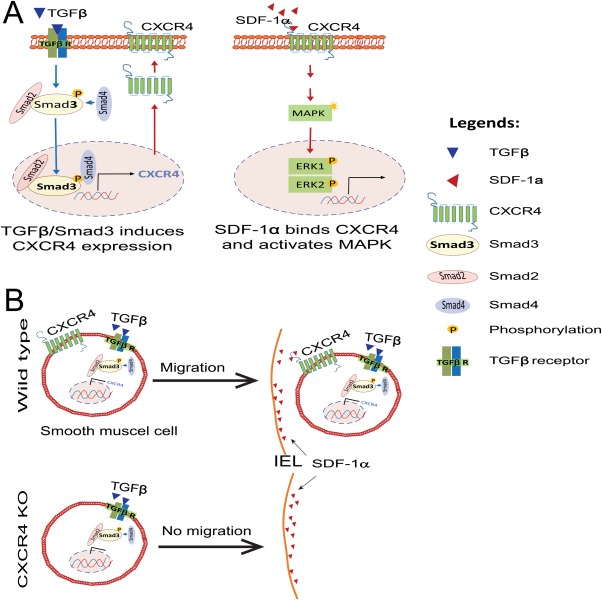
CXCR4 is a stem/progenitor cell surface receptor specific for the cytokine stromal cell-derived factor-1 (SDF-1α). There is evidence that bone marrow-derived CXCR4-expressing cells contribute to intimal hyperplasia (IH) by homing to the arterial subintima which is enriched with SDF-1α. We have previously found that transforming growth factor-β (TGFβ) and its signaling protein Smad3 are both upregulated following arterial injury and that TGFβ/Smad3 enhances the expression of CXCR4 in vascular smooth muscle cells (SMCs). It remains unknown, however, whether locally induced CXCR4 expression in SM22 expressing vascular SMCs plays a role in neointima formation. Here, we investigated whether elevated TGFβ/Smad3 signaling leads to the induction of CXCR4 expression locally in the injured arterial wall, thereby contributing to IH. We found prominent CXCR4 upregulation (mRNA, 60-fold; protein, 4-fold) in TGFβ-treated, Smad3-expressing SMCs. Chromatin immunoprecipitation assays revealed a specific association of the transcription factor Smad3 with the CXCR4 promoter. TGFβ/Smad3 treatment also markedly enhanced SDF-1α-induced ERK1/2 phosphorylation as well as SMC migration in a CXCR4-dependent manner. Adenoviral expression of Smad3 in balloon-injured rat carotid arteries increased local CXCR4 levels and enhanced IH, whereas SMC-specific depletion of CXCR4 in the wire-injured mouse femoral arterial wall produced a 60% reduction in IH. Our results provide the first evidence that upregulation of TGFβ/Smad3 in injured arteries induces local SMC CXCR4 expression and cell migration, and consequently IH. The Smad3/CXCR4 pathway may provide a potential target for therapeutic interventions to prevent restenosis. Stem Cells 2016;34:2744-2757.

Keywords: CXCR4/SDF-1α; TGFβ/Smad3; intimal hyperplasia; smooth muscle cell migration; smooth muscle cell specific CXCR4 knockout.


Figures

Figure 1
Figure 1
TGFβ/Smad3 stimulates CXCR4 expression in cultured rat aortic smooth muscle cells (SMCs). (A): Quantitative RT‐PCR. mRNA levels of CXCR4 were determined from SMCs infected with AdGFP (control) or AdSmad3 followed by treatment with solvent or TGFβ1 for 6 hour. Error bar represents SEM.*p < .05 compared with AdGFP control; **p < .05 compared with all other conditions; n = 3. (B): Quantitative RT‐PCR. mRNA levels of CXCR4 were determined with SMCs transfected with scrambled siRNA (control) or Smad3 siRNA followed by treatment with TGFβ1 for 6 hour. *p < .05 compared with solvent (0 ng/ml) in control siRNA group; **p < .01 compared with all other conditions in control siRNA group; #p < .05 compared with 2.5 ng/ml TGFβ1 treatment in control siRNA group; ##p < .05 compared with 5 ng/ml TGFβ1 treatment in control siRNA group; n = 3. (C): Western blotting. Protein levels of CXCR4 were assessed from SMCs infected with AdGFP or AdSmad3 followed by treatment with solvent or TGFβ1 (5 ng/ml) for 24 hour. *p < .05 compared with AdGFP control; **p < .05 compared with all other conditions; n = 3. (D): Immunocytochemistry. CXCR4 expression on SMCs was visualized (red) using SMCs treated with AdGFP or AdSmad3 followed by treatment with TGFβ1 (5 ng/ml) for 24 hour. Nuclei were stained blue by 4′,6‐diamidino‐2‐phenylindole (DAPI). Scale bar = 20 µm. Image magnification: 200×. Abbreviation: TGFβ, transforming growth factor‐β.
Figure 2
Figure 2
TGFβ/Smad3 treatment increases Smad3 binding to the CXCR4 promoter. (A): Schematic illustration of the rat CXCR4 promoter depicting the location of Smad binding element (SBE) and chromatin immunoprecipitation (ChIP) primer sets relative to the transcription start site. ChIP was performed using Smad3 antibody or normal IgG as described in the Methods section. Quantitative RT‐PCR was performed with precipitated DNA using primer sets flanking SBE amplifying the CXCR4 promoter proximal region. A primer set amplifying the distal region of CXCR4 promoter was used as a control. *p < .05, compared with AdGFP control; **p < .05, compared with AdGFP, AdGFP +TGFβ or AdSmad3; n = 3. (B): Electrophoretic mobility shift assay (EMSA) was performed as described in Methods. A synthetic biotinylated oligo containing the SBE from the CXCR4 promoter was used to react with nuclear protein extracts from AdSmad3‐infected and then TGFβ‐treated smooth muscle cells (SMCs). Shifted bands were detected with streptavidin‐conjugated horseradish peroxidase. The same DNA oligo without biotin (unlabeled oligo) was used to compete with the biotinylated oligo for binding with Smad3. (C): EMSA was performed with increasing amounts of unlabeled oligo to compete with the biotinylated oligo for binding with Smad3. (D): EMSA in the presence of a Smad3 antibody showed super shift. (E): A synthetic biotinylated oligo containing the SBE of the CXCR4 promoter was incubated with nuclear protein extracts from AdSmad3 infected and TGFβ1‐treated SMCs. Protein bound to biotinylated oligo was precipitated with avidin beads. Western blot was performed with a Smad3 antibody. A biotinylated oligo containing a mutated SBE or unlabeled SBE oligo were used as controls. Abbreviation: TGFβ, transforming growth factor‐β.
Figure 3
Figure 3
TGFβ/Smad3 treatment stimulates CXCR4‐dependent smooth muscle cell (SMC) migration toward SDF‐1α. (A): Binding of biotin‐SDF‐1α to CXCR4 expressed on the unpermeabilized SMC surface was assayed via streptavidin‐conjugated HRP, as described in Methods. Prior to the addition of biotin‐SDF‐1α, SMCs were infected with AdGFP (control) or AdSmad3 and then treated with solvent or TGFβ1 for 24 hour. Error bar represents SEM; *p < .05 compared with AdGFP control; **p < .05 compared with all other three conditions (n = 3). (B): Migration assay was performed as described in Methods. SMCs were infected with AdGFP (control) or AdSmad3 followed by treatment with solvent or TGFβ1 for 24 hour, and then transferred to a Boyden Transwell insert. AMD3100 (10 μM) or solvent were added to the upper chamber. Sixty minutes later, SDF‐1α was added to the lower chamber (100 ng/ml). After 4 hour, the cells that migrated through the Transwell membrane were counted. *p < .05 compared with AdGFP control; **p < .05 compared with all other conditions; #p < .05 compared with AdGFP+TGFβ treated group without AMD3100; ##p < .05 compared with AdSmad3 treated group without AMD3100; ###p < .05 compared with AdSmad3+TGFβ treated group without AMD3100; n = 3. Abbreviation: SDF‐1, stromal cell‐derived factor‐1; TGFβ, transforming growth factor‐β.
Figure 4
Figure 4
TGFβ/Smad3 treatment stimulates SDF‐1α‐induced, CXCR4‐dependent ERK phosphorylation in smooth muscle cells (SMCs). AdSmad3 infected and TGFβ1 (5 ng/ml) treated SMCs were stimulated with SDF‐1α and ERK phosphorylation was detected by Western blotting. (A‐D): Dose response of SDF1α induced (10 min) ERK phosphorylation in SMCs treated with AdGFP (A), AdGFP +TGFβ1 (B), AdSmad3 (C), and AdSmad3+TGFβ1 (D), respectively. *p < .05 compared with 0 ng/ml; **p < .01 compared with 0 ng/ml; n = 3. (E): AdSmad3 infected and TGFβ1 (5 ng/ml) treated SMCs were stimulated with SDF‐1α (100 ng/ml) for indicated time and ERK phosphorylation was detected by Western blotting. *p < .05 compared with 0 min; **p < .01 compared with 0 min; n = 3. (F): SDF‐1α‐induced (10 min, 100 ng/ml) ERK phosphorylation in SMCs without or with AMD3100 (10 µM). *p < .05 compared with AdGFP in control group; **p < .05 compared with all other conditions in control group; #p < .05 compared with AdGFP+TGFβ in control group; ##p < .05 compared with AdSmad3 in control group; ###p < .05 compared with AdSmad3+TGFβ in control group; n = 3. (G): SMCs were isolated from aorta of wild type or CXCR4 conditional knockout mice and treated with control (solvent, 4 mM HCl) or TGFβ1 (5 ng/ml) for 24 hour. SDF‐1α‐induced (10 min; 100 ng/ml) ERK phosphorylation was analyzed by Western blotting. *p < .05 compared with all other conditions, n = 3. (H): SMCs were infected with AdGFP (control) or AdSmad3 followed by treatment with TGFβ1 (5 ng/ml) for 24 hour, and then transferred to a Boyden Transwell insert. Cells were incubated with solvent or U0126 (5 µM) or PD989059 (10 µm) for 60 min, and then 100 ng/ml SDF‐1α was added to the lower chamber. The cells that migrated through the Transwell insert membrane after 4 hour were counted. *p < .05 compared with AdGFP; **p < .05 compared with AdSmad3+TGFβ without an inhibitor; n = 3. Abbreviations: SDF‐1, stromal cell‐derived factor‐1; TGFβ, transforming growth factor‐β.
Figure 5
Figure 5
Enhanced Smad3 expression up‐regulates CXCR4 production in balloon‐injured rat carotid arteries. Balloon angioplasty was performed in rat carotid arteries followed by infusion of AdGFP or AdSmad3 (2.5 × 10⁹ plaque‐forming units) for 20 minutes. Uninjured carotid arteries were used as control. Carotid arteries were retrieved at the indicated time points (3, 7, or 14 days) for preparation of cross sections. (A and B): Immunostaining was performed to detect CXCR4 (red) and DAPI was used to stain nuclei (blue). Dashed lines define the media layer; arrowheads mark internal elastic lamina (IEL). (C): Relative fluorescent intensity was measured using ImageJ software. Each bar is a mean ± SEM (n= 3‐5); *p < .05 compared with AdGFP control; **p < .05 compared with all other conditions. (D): Western blot was performed on proteins extracted from uninjured or injured with AdGFP or AdSmad3 infused carotid arteries 3 days after angioplasty. (E): Immunostaining of Smad3 on sections from uninjured, or injured with AdGFP or AdSmad3 infused carotid artery. Dashed lines define the media layer; arrowheads mark IEL. (F): RT‐PCR for CXCR4 levels after vascular injury. *p < .05 compared with uninjured; **p < .05 compared with uninjured or injured with AdGFP infused; n = 3. (G): Immunostaining of SDF‐1α on sections of carotid arteries on 3, 7, or 14 days after injury. Dashed lines define the media layer; arrowheads mark IEL. (H) and (J) are immunochemistry of Ki67 or Smad3 in injured sections of carotid arteries at the indicated time points (3, 7, or 14 days). (I) and (K) are magnified views of the boxed regions in H and J, respectively. Scale bar = 30 µm.
Figure 6
Figure 6
Selective knockout of CXCR4 in smooth muscle cells (SMCs) reduces intimal hyperplasia (IH) in wire‐injured mouse femoral arteries. SM22‐driven conditional CXCR4 knockout model was created and wire injury was performed in wild type and CXCR4 knockout mice, as described in Methods. (A–E): Representative H&E stained femoral arterial sections in uninjured (A), injured wild type (B) or injured knockout mice (C). (D) and (E) are enlarged views of the boxed areas in (B) and (C). (FJ): CXCR4 immunostaining in uninjured (F), injured wild type (G), or injured knockout mice (H). (I) and (J) are enlargement of the boxed regions from (G) and (H). (K‐M): SDF‐1α immunostaining in uninjured (K), injured wild type (L), or injured knockout mice (M). (N–O): SMA immunostaining in injured wild type (N) or injured knockout mice (O). (P–R): Smad3 immunostaining in uninjured (P), injured wild type (Q), or injured knockout mice (R). (S): IH (intima/media area ratio), lumen area, and media area were quantified as described in Methods. *p < .05 compared to wild type; n = 12‐13. Scale bar = 50 µm. Abbreviation: SDF‐1, stromal cell‐derived factor‐1.
Figure 7
Figure 7
A schematic model showing the role of TGFβ/Smad3 regulated CXCR4 expression in SMC migration toward SDF‐1α. (A): TGFβ/Smad3 stimulates CXCR4 expression while SDF‐1α activates MAPK through CXCR4. (B): Elevated SDF‐1α in the subintima of injured artery attracts SMCs from wildtype mice but not from CXCR4 knockout mice. Abbreviations: SDF‐1, stromal cell‐derived factor‐1; SMCs, smooth muscle cells; TGFβ, transforming growth factor‐β.

Similar articles

Cited by

References

    1. Mills B, Robb T, Larson DF. Intimal hyperplasia: slow but deadly. Perfusion 2012;27:520–528. - PubMed
    1. Suwanabol PA, Kent KC, Liu B. TGF‐β and restenosis revisited: a smad link. J Surg Res 2011;167:287–297. - PMC - PubMed
    1. Akhurst RJ, Hata A. Targeting the TGFβ signaling pathway in disease. Nat Rev Drug Discov 2012;11:790–811. - PMC - PubMed
    1. Tsai S, Butler J, Rafii S, et al. The role of progenitor cells in the development of intimal hyperplasia. J Vasc Surg 2009;49:502–510. - PMC - PubMed
    1. Edlin RS, Tsai S, Yamanouchi D, et al. Characterization of primary and restenotic atherosclerotic plaque from the superficial femoral artery: potential role of Smad3 in regulation of SMC proliferation. J Vasc Surg 2009;49:1289–1295. - PMC - PubMed

Publication types

MeSH terms