Review
. 2016 Sep 1;23(9):1417-27.
doi: 10.1038/cdd.2016.52. Epub 2016 Jun 24.

The coming of age of the mitochondria-ER contact: a matter of thickness

Affiliations
Review

The coming of age of the mitochondria-ER contact: a matter of thickness

M Giacomello et al. Cell Death Differ. .

Abstract

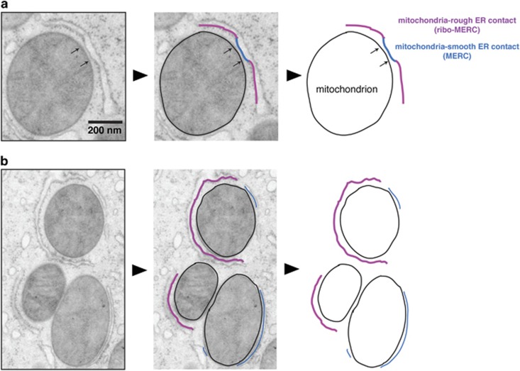
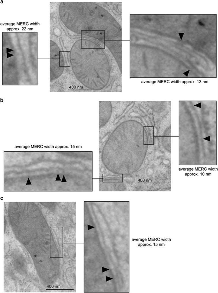
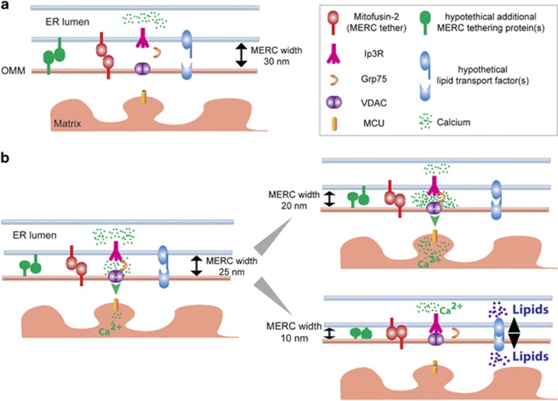
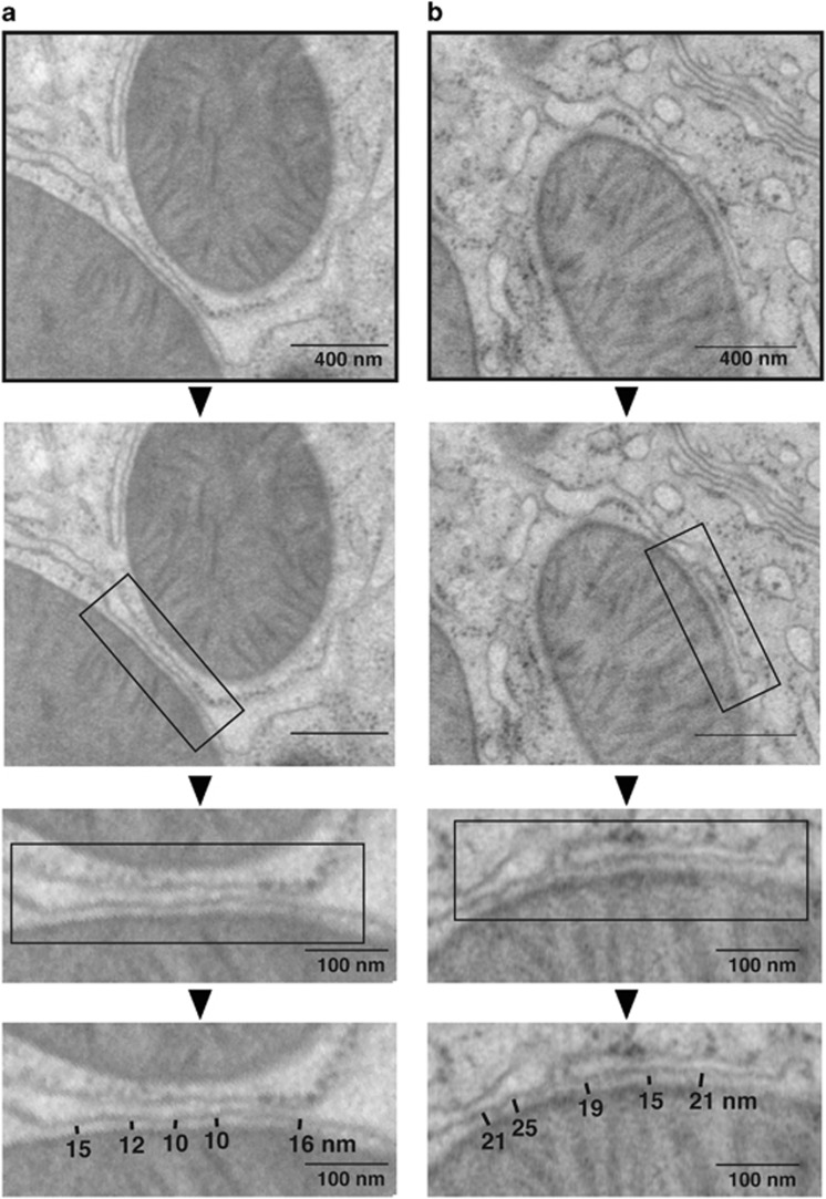
The sites of near-contact between the mitochondrion and the endoplasmic reticulum (ER) have earned a lot of attention due to their key role in the maintenance of lipid and calcium (Ca(2+)) homeostasis, in the initiation of autophagy and mitochondrial division, and in sensing metabolic shifts. At these sites, typically called MAMs (mitochondria-associated ER membranes) or MERCs (mitochondria-ER contacts), the organelles juxtapose at a distance that can range from ~10 to ~50 nm. The multifunctional role of this subcellular compartment is puzzling; further, recent studies have shown that mitochondria-ER contacts are highly plastic structures that remodel upon metabolic transitions and that their activity in controlling lipid homeostasis could be involved in Alzheimer's disease pathogenesis. This review aims at integrating the functions of this subcellular compartment to its most characterizing and unexplored structural parameter, their 'thickness': that is, the width of the cleft that separates the cytosolic face of the outer mitochondrial membrane from that of the ER. We describe and discuss the reasons why the thickness of a MERC should be considered a regulated structural parameter of the cell that defines and controls its function. Further, we propose a MERC classification that will help organize the expanding field of MERCs biology and of their role in cell physiology and human disease.


Figures

Figure 1
Figure 1
Mitochondria form sites of contact with the smooth ER (MERCs) and with the ribosome-containing rough ER (ribo-MERCs). These two types of structures can exist as part of a single unit (a) or as separate entities (b). MERCs are the most-studied ones because they are responsible for ions and lipid transfer between the two organelles; ribo-MERCs, instead, have been so far poorly studied and their function is still unknown. Both types of mitochondria–ER contacts typically extend over several hundreds nanometer in length. In mouse liver hepatocytes (shown here), the vast majority of the MERCs are characterized by having the two organelles juxtaposed at a constant distance that varies between ~10 and ~25 nm for the MERCs, and ~50 to ~80 nm for the ribo-MERCs. The length and the thickness of the MERC change during hepatic metabolic shifts, indicating that they are regulated structural parameters of the cell. The reason behind the structural plasticity of the MERCs remains unknown, but it could be a key part of the mitochondrial adaptive response that contributes to build the metabolic flexibility of the cell
Figure 2
Figure 2
MERCs of various thicknesses are filled by electron-dense structures. Cryo-EM images from mouse hepatocytes showing MERC clefts of different average width decorated by electron-dense strcutures (a–c; arrows); these structures are likely formed by proteins that tether the two membranes together and that form the complexes that drive the function of the MERC
Figure 3
Figure 3
Model depicting how changes in the distance that separates the mitochondria from the ER could regulate MERCs function and/or level of activity. MERCs are dynamic structures; the width of the cleft changes depending on the metabolic state of the cell. This model shows how a too wide or too narrow distance between the organelles could affect the assembly of the complexes that drive calcium and lipid fluxes. A width of 30 nm is likely to result in a functionally dormant MERC (a); based on Fick and Einstein's diffusion laws, a cleft 20 nm wide would support higher rates and volumes of calcium uptake than one that is 25-nm wide (b); however, a cleft of 10 nm would impair this process, due to steric hindrance of the proteins that form the complex that regulates its flux from the ER to the mitochondrion. Instead, a 10-nm wide MERC cleft could allow the formation of the proteins that mediate lipid transfer (c), which likely requires the tunneling of a lipid through hydrophobic channels formed by protein complexes anchored to the two juxtaposed membranes
Figure 4
Figure 4
The width of the MERC cleft is rather uniform, but varies from MERC to MERC. Representative cryo-EM images of mouse hepatocytes showing a MERC with an average constant width of ≈12 nm (a) and ≈20 nm (b). As the distance between the organelles is a regulated structural parameter of the cell and is subjected to Fick and Einstein's diffusion laws, it is conceivable that the two MERCs shown here could have different function and/or level of activity

References

    1. Copeland DE, Dalton AJ. An association between mitochondria and the endoplasmic reticulum in cells of the pseudobranch gland of a teleost. J Biophys Biochem Cytol 1959; 5: 393–396. - PMC - PubMed
    1. Montisano DF, Cascarano J, Pickett CB, James TW. Association between mitochondria and rough endoplasmic reticulum in rat liver. Anat Rec 1982; 203: 441–450. - PubMed
    1. Pickett CB, Montisano D, Eisner D, Cascarano J. The physical association between rat liver mitochondria and rough endoplasmic reticulum. I. Isolation, electron microscopic examination and sedimentation equilibrium centrifugation analyses of rough endoplasmic reticulum-mitochondrial complexes. Exp Cell Res 1980; 128: 343–352. - PubMed
    1. Sood A, Jeyaraju VD, Prudent J, Caron A, Lemieux P, McBride HM et al. A Mitofusin-2-dependent inactivating cleavage of Opa1 links changes in mitochondria cristae and ER contacts in the postprandial liver. Proc Natl Acad Sci USA 2014; 111: 16017–16022. - PMC - PubMed
    1. Bravo R, Vicencio JM, Parra V, Troncoso R, Munoz JP, Bui M et al. Increased ER-mitochondrial coupling promotes mitochondrial respiration and bioenergetics during early phases of ER stress. J Cell Sci 2011; 124: 2143–2152. - PMC - PubMed

Publication types

Grants and funding

LinkOut - more resources