. 2016 Nov;31(11):1920-1929.
doi: 10.1002/jbmr.2892. Epub 2016 Oct 24.

Identification of Senescent Cells in the Bone Microenvironment

Affiliations

Identification of Senescent Cells in the Bone Microenvironment

Joshua N Farr et al. J Bone Miner Res. 2016 Nov.

Abstract

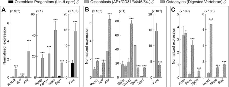
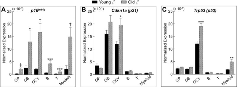
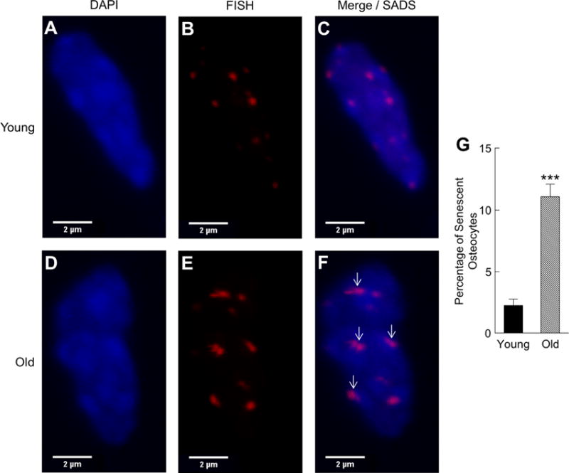
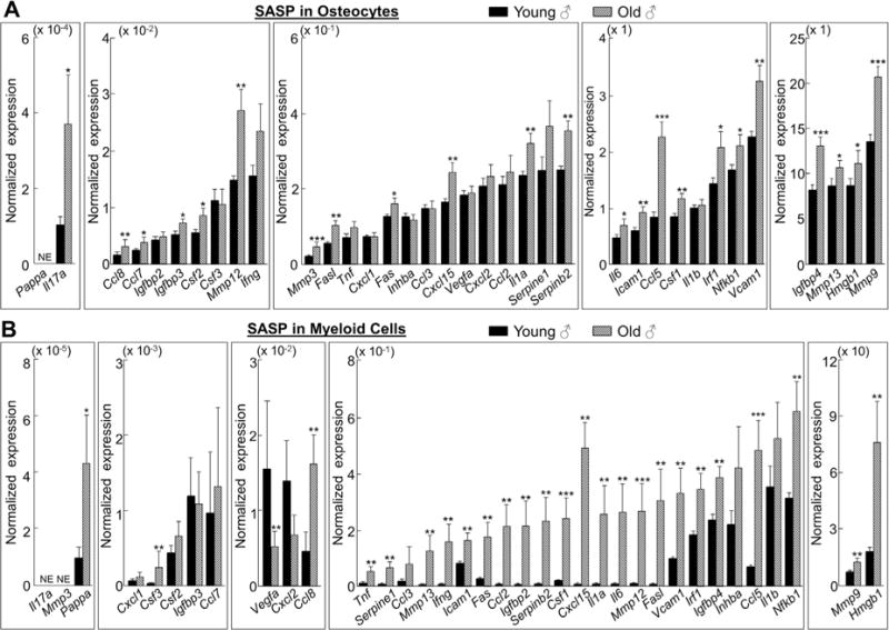
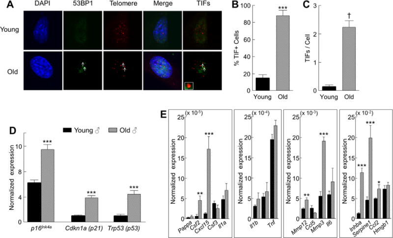
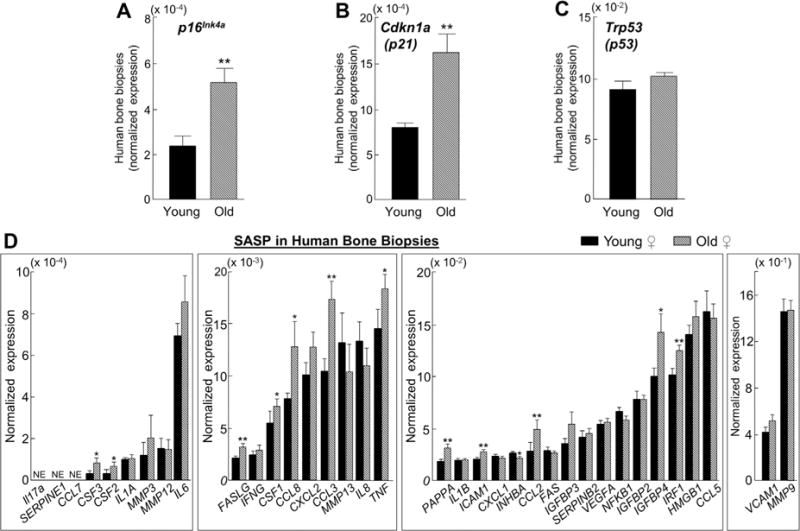
Cellular senescence is a fundamental mechanism by which cells remain metabolically active yet cease dividing and undergo distinct phenotypic alterations, including upregulation of p16Ink4a , profound secretome changes, telomere shortening, and decondensation of pericentromeric satellite DNA. Because senescent cells accumulate in multiple tissues with aging, these cells and the dysfunctional factors they secrete, termed the senescence-associated secretory phenotype (SASP), are increasingly recognized as promising therapeutic targets to prevent age-related degenerative pathologies, including osteoporosis. However, the cell type(s) within the bone microenvironment that undergoes senescence with aging in vivo has remained poorly understood, largely because previous studies have focused on senescence in cultured cells. Thus in young (age 6 months) and old (age 24 months) mice, we measured senescence and SASP markers in vivo in highly enriched cell populations, all rapidly isolated from bone/marrow without in vitro culture. In both females and males, p16Ink4a expression by real-time quantitative polymerase chain reaction (rt-qPCR) was significantly higher with aging in B cells, T cells, myeloid cells, osteoblast progenitors, osteoblasts, and osteocytes. Further, in vivo quantification of senescence-associated distension of satellites (SADS), ie, large-scale unraveling of pericentromeric satellite DNA, revealed significantly more senescent osteocytes in old compared with young bone cortices (11% versus 2%, p < 0.001). In addition, primary osteocytes from old mice had sixfold more (p < 0.001) telomere dysfunction-induced foci (TIFs) than osteocytes from young mice. Corresponding with the age-associated accumulation of senescent osteocytes was significantly higher expression of multiple SASP markers in osteocytes from old versus young mice, several of which also showed dramatic age-associated upregulation in myeloid cells. These data show that with aging, a subset of cells of various lineages within the bone microenvironment become senescent, although senescent myeloid cells and senescent osteocytes predominantly develop the SASP. Given the critical roles of osteocytes in orchestrating bone remodeling, our findings suggest that senescent osteocytes and their SASP may contribute to age-related bone loss. © 2016 American Society for Bone and Mineral Research.

Keywords: AGING; ANIMAL MODELS; CELL/TISSUE SIGNALING; OSTEOCYTES.


Conflict of interest statement

All authors state that they have no conflicts of interest.

Figures

Fig. 1
Fig. 1
Comparisons of key osteoblast lineage markers among osteoblast progenitors, osteoblasts, and osteocytes. (A) The normalized mRNA expression of osteoblast genes is significantly higher in osteoblasts (AP+/CD31/34/45/54− cells) as compared with osteoblast progenitors (Lin−/Lepr+ cells). (B) Comparisons of normalized mRNA expression between osteoblasts (AP+/CD31/34/45/54− cells) and osteocytes (digested vertebrae). (C) The normalized mRNA expression of osteocyte genes is significantly higher in osteocytes (digested vertebrae) as compared with osteoblasts (AP+/CD31/34/45/54− cells). All three cell populations were derived from young (age 6 months) male mice (n = 12). Data are presented as mean ± SE. ***p < 0.001.
Fig. 2
Fig. 2
Cells in the bone microenvironment from old mice express higher levels of the senescence biomarker p16Ink4a. In vivo age-associated changes in normalized mRNA expression of senescence effectors (A) p16Ink4a, (B) p21, and (C) p53 are shown for OP (osteoblast progenitors; Lin−/Lepr+ cells), OB (osteoblasts; AP+/CD31/34/45/54− cells), OCY (osteocytes; digested vertebrae), B (B cells; CD19+/CD14/15/3ε− cells), T (T cells; CD3ε+/CD14/15/19− cells), and myeloid (myeloid cells; CD14+ cells), all rapidly isolated from young (age 6 months; n = 12) and old (age 24 months; n = 10) male mice (without in vitro culture). Data are presented as mean ± SE. *p < 0.05; **p < 0.01; ***p < 0.001; p < 0.0001; p < 0.00001.
Fig. 3
Fig. 3
A subset of osteocytes from old mice display marked distension of pericentromeric satellite DNA in vivo. Senescence-associated distension of satellites (SADS, see arrows [in F]) in osteocytes from young (age 6 months; AC) versus old (age 24 months; DF) male cortical bone diaphyses (n = 4 per group; magnification ×100). (G) Quantification of the percentage of senescent osteocytes in young versus old male mice based on a cut-off of ≥4 SADS per cell (see Supplementary Methods for determination of this cut-off). ***p < 0.001.
Fig. 4
Fig. 4
Osteocytes and myeloid cells from old chronologically aged mice develop the senescence-associated secretory phenotype (SASP). (A) Osteocytes and (B) myeloid cells were rapidly isolated from young (age 6 months; n = 12) and old (age 24 months; n = 10) male mice (without in vitro culture). In vivo age-associated changes in normalized mRNA expression of 36 established SASP components are shown. Results are expressed as mean ± SE. NE = not expressed (Cycle threshold [Ct] values >35 in both young and old samples). *p < 0.05; **p < 0.01; ***p < 0.001.
Fig. 5
Fig. 5
Primary osteocytes from old chronologically aged mice have dysfunctional telomeres and maintain their senescence-associated secretory phenotype (SASP) when put into culture. (A) Telomere dysfunction-induced foci (TIFs), co-localization of 53BP1 (a DNA damage protein), and telomeric DNA are shown for primary osteocytes (arrows) from old (age 24 months) male mice; inset shows magnification of representative TIF (magnification× 630). (B) Percentage of TIF+ cells and (C) TIFs per cell in primary cultured osteocytes from old (age 24 months; n = 3) versus young (age 4 months; n = 3) male mice. In vivo age-associated changes in normalized mRNA expression of (D) senescence effectors and (E) SASP factors in male primary osteocytes after 7 days in culture. Data are presented as mean ± SE. *p < 0.05; **p < 0.01; ***p < 0.001; p < 0.0001.
Fig. 6
Fig. 6
Expression levels of senescence effectors and senescence-associated secretory phenotype (SASP) markers are higher with aging in human bone biopsies. Small needle bone biopsies were obtained from the posterior iliac crest of 10 young (mean ± SD; 27 ± 3 years) and 10 old (mean ± SD; 78 ± 6 years) healthy female volunteers and the normalized expression of (AC) senescence and (D) SASP genes was measured in vivo using rt-qPCR. Data are presented as mean ± SE. NE = not expressed (Cycle threshold [Ct] values >35 in both young and old samples). *p < 0.05; **p < 0.01.

Comment in

References

    1. Goldman DP, Cutler D, Rowe JW, et al. Substantial health and economic returns from delayed aging may warrant a new focus for medical research. Health Aff (Millwood) 2013;32(10):1698–1705. - PMC - PubMed
    1. Tchkonia T, Zhu Y, Van Deuersen J, Campisi J, Kirkland JL. Cellular senescence and the senescent secretory phenotype: therapeutic opportunities. J Clin Invest. 2013;123:966–72. - PMC - PubMed
    1. van Deursen JM. The role of senescent cells in ageing. Nature. 2014;509:439–46. - PMC - PubMed
    1. Childs BG, Durik M, Baker DJ, van Deursen JM. Cellular senescence in aging and age-related disease: from mechanisms to therapy. Nat Med. 2015;21(12):1424–35. - PMC - PubMed
    1. Swanson EC, Manning B, Zhang H, Lawrence JB. Higher-order unfolding of satellite heterochromatin is a consistent and early event in cell senescence. J Cell Biol. 2013;203(6):929–42. - PMC - PubMed

Publication types