. 2016 Jun 24:13:111.
doi: 10.1186/s12985-016-0567-6.

Antiviral activity of Lactobacillus reuteri Protectis against Coxsackievirus A and Enterovirus 71 infection in human skeletal muscle and colon cell lines

Affiliations

Antiviral activity of Lactobacillus reuteri Protectis against Coxsackievirus A and Enterovirus 71 infection in human skeletal muscle and colon cell lines

Lei Yin Emily Ang et al. Virol J. .

Erratum in

Abstract

Background: Recurrence of hand, foot and mouth disease (HFMD) pandemics continues to threaten public health. Despite increasing awareness and efforts, effective vaccine and drug treatment have yet to be available. Probiotics have gained recognition in the field of healthcare worldwide, and have been extensively prescribed to babies and young children to relieve gastrointestinal (GI) disturbances and diseases, associated or not with microbial infections. Since the faecal-oral axis represents the major route of HFMD transmission, transient persistence of probiotic bacteria in the GI tract may confer some protection against HFMD and limit transmission among children.

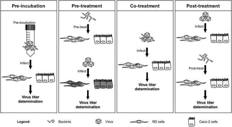
Methods: In this work, the antiviral activity of two commercially available probiotics, namely Lactobacillus reuteri Protectis (L. reuteri Protectis) and Lactobacillus casei Shirota (L. casei Shirota), was assayed against Coxsackieviruses and Enterovirus 71 (EV71), the main agents responsible for HFMD. In vitro infection set-ups using human skeletal muscle and colon cell lines were designed to assess the antiviral effect of the probiotic bacteria during entry and post-entry steps of the infection cycle.

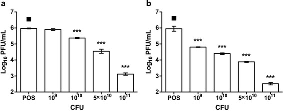
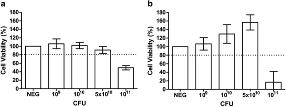
Results: Our findings indicate that L. reuteri Protectis displays a significant dose-dependent antiviral activity against Coxsackievirus type A (CA) strain 6 (CA6), CA16 and EV71, but not against Coxsackievirus type B strain 2. Our data support that the antiviral effect is likely achieved through direct physical interaction between bacteria and virus particles, which impairs virus entry into its mammalian host cell. In contrast, no significant antiviral effect was observed with L. casei Shirota.

Conclusions: Should the antiviral activity of L. reuteri Protectis observed in vitro be translated in vivo, such probiotics-based therapeutic approach may have the potential to address the urgent need for a safe and effective means to protect against HFMD and limit its transmission among children.

Keywords: Coxsackievirus; Enterovirus 71; Foot and mouth disease; Hand; Lactobacillus reuteri; Probiotics.


Figures

Fig. 1
Fig. 1
Cell viability in the presence of live L. reuteri Protectis bacteria. Different concentrations of L. reuteri Protectis bacteria were added to RD cells (a) and Caco-2 cells (b) and incubated for 1 h, then washed with PBS thrice before 50 μg/ml gentamicin-supplemented maintenance media was added to the cells. Alamar blue assay was performed at 24 h post-treatment according to the manufacturer’s instructions. Data are expressed as the mean ± standard deviation of technical triplicates. Two biological repeats were conducted. One representative is shown
Fig. 2
Fig. 2
Schematic diagram of experimental in vitro set-ups. In the pre-incubation set-up, live bacteria and virus were pre-incubated for 1 h at 37 °C, before being incubated with the mammalian cells for 1 h. In the pre-treatment set-up, the mammalian cells were incubated with live bacteria for 1 h, washed with PBS and infected with virus for 1 h. In the co-treatment set-up, live bacteria were added to the cells at the same time of virus infection for 1 h. In the post-treatment set-up, the mammalian cells were infected with virus for 1 h, then washed with PBS and incubated with live bacteria for another hour. In all four set-ups, 50 μg/ml gentamicin-supplemented maintenance media was eventually added to the cell monolayers. Sample supernatants were harvested 12 or 24 h post-infection and plaque assay was performed using RD cells to determine the viral titer
Fig. 3
Fig. 3
Antiviral effect of L. reuteri Protectis. Pre-incubation, pre-treatment, co-treatment and post-treatment setups were performed as detailed in Fig. 1. Virus titers in the supernatant of CA6- a, CA16- b, EV71- c and CB2- d infected RD cells and Caco-2 cells were determined by standard plaque assay in RD cells. A one-way ANOVA test with Dunnett’s posttest was performed (* p < 0.05, ** p < 0.005, *** p < 0.0005). Data are expressed as the mean ± standard deviation of technical triplicates. Two biological repeats were conducted. One representative is shown
Fig. 4
Fig. 4
Immunostaining of RD cells infected with (L. reuteri + CA16) mixture. a L. reuteri Protectis bacteria (1011 CFU) were co-incubated with CA16 (105 PFU) for 1 h in 37 °C prior to adding the mixture onto RD cells (105 cells) for another 1 h. A control with RD cells infected with CA16 only was also performed. The monolayers were then washed thrice before methanol fixation and immunostained with anti-CA16 and anti-beta actin antibodies. Nuclei were also stained with DAPI. Images were taken using Olympus IX81 microscope. b Corrected total cell fluorescence (CTCF) (viral signal) was calculated from each cell originated from three random microscopic views. Statistical analysis was performed using Mann–Whitney U test (****, p < 0.0001)
Fig. 5
Fig. 5
Virus-bacteria binding assay. Different quantities of live L. reuteri Protectis bacteria were incubated with a fixed amount of EV71, CA6, CA16 or CB2 virus particles for 1 h. The mixtures were then centrifuged and filtered to obtain free virus in the supernatant. Virus titers were determined by plaque assay using RD cells. A one-way ANOVA test with Dunnett’s posttest was performed (* p < 0.05, ** p < 0.005, *** p < 0.0005). Data are expressed as the mean ± standard deviation of technical triplicates. Two biological repeats were conducted. One representative is shown
Fig. 6
Fig. 6
Antiviral effect of formaldehyde-inactivated L. reuteri Protectis. Formalin-fixed L. reuteri Protectis bacteria were bacteria were pre-incubated with CA6 or CA16 virus according to the pre-incubation set-up with Caco-2 cells. Virus titers in the supernatant of CA6- (a) and CA16- (b) infected cells were determined by standard plaque assay in RD cells. A one-way ANOVA test with Dunnett’s posttest was performed (* p < 0.05, ** p < 0.005, *** p < 0.0005). Data are expressed as the mean ± standard deviation of technical triplicates. Two biological repeats were conducted. One representative is shown
Fig. 7
Fig. 7
Cell viability in the presence of live L. casei Shirota. Different concentrations of bacteria were added to RD (a) and Caco-2 (b) cells as indicated and incubated for 1 h, then washed with 1xPBS thrice before 50 μg/ml gentamicin-supplemented maintenance media was added to the cells. Alamar blue assay was performed at 24 h post-treatment according to the manufacturer’s instructions. Data are expressed as the mean ± standard deviation of technical triplicates. Two biological repeats were conducted. One representative is shown
Fig. 8
Fig. 8
Antiviral effect of L. casei Shirota. Pre-incubation, pre-treatment, co-treatment and post-treatment setups were performed as detailed in Fig. 1. Virus titers in the supernatant of CA16- (a) and EV71- (b) infected RD cells and Caco-2 cells were determined by standard plaque assay in RD cells. A one-way ANOVA test with Dunnett’s posttest was performed (* p < 0.05, ** p < 0.005, *** p < 0.0005). Data are expressed as the mean ± standard deviation of technical triplicates. Two biological repeats were conducted. One representative is shown

References

    1. Bruu AL: Enteroviruses: Polioviruses, coxsackieviruses, echoviruses, and newer enteroviruses. A Practical Guide to Clinical Virology, Second Edition 2003;44-45. doi:10.1002/0470857285.ch6
    1. Hirata O, Ishikawa N, Mizoguchi Y, Nakamura K, Kobayashi M. A case of neonatal coxsackie B2 meningo-encephalitis in which serial magnetic resonance imaging findings reveal the development of lesions. Neuropediatrics. 2011;42:156–8. doi: 10.1055/s-0031-1285876. - DOI - PubMed
    1. Wang SM, Liu CC. Update of enterovirus 71 infection: epidemiology, pathogenesis and vaccine. Expert Rev Anti Infect Ther. 2014;12:447–56. doi: 10.1586/14787210.2014.895666. - DOI - PubMed
    1. Huang H-I, Weng K-F, Shih S-R. Viral and host factors that contribute to pathogenicity of enterovirus 71. Future Microbiol. 2012;7:467–79. doi: 10.2217/fmb.12.22. - DOI - PubMed
    1. Mao Q, Wang Y, Yao X, Bian L, Wu X, Xu M, Liang Z. Coxsackievirus A16: epidemiology, diagnosis, and vaccine. Hum Vaccin Immunother. 2014;10:360–7. doi: 10.4161/hv.27087. - DOI - PMC - PubMed

Publication types