. 2016 Jun 24;17(1):21.
doi: 10.1186/s12865-016-0160-1.

Immunoregulatory effects triggered by immunobiotic Lactobacillus jensenii TL2937 strain involve efficient phagocytosis in porcine antigen presenting cells

Affiliations

Immunoregulatory effects triggered by immunobiotic Lactobacillus jensenii TL2937 strain involve efficient phagocytosis in porcine antigen presenting cells

Kohichiro Tsukida et al. BMC Immunol. .

Abstract

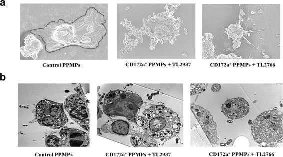
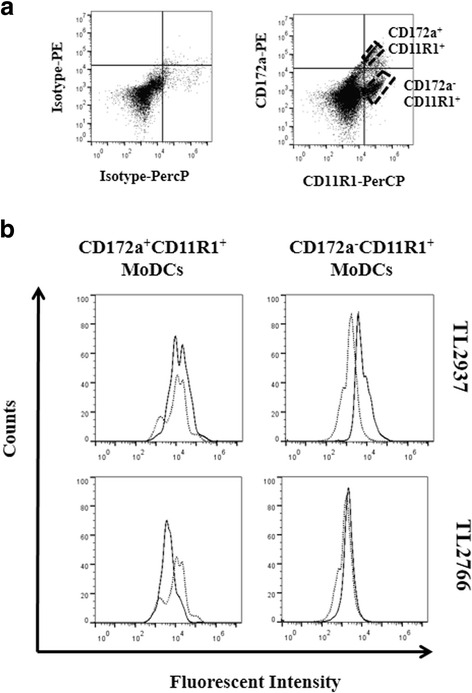
Background: Immunobiotic Lactobacillus jensenii TL2937 modulates porcine mononuclear phagocytes from Peyer's patches (PPMPs) and induces a differential production of pro- and anti-inflammatory cytokines in response to Toll-like receptor (TLR)-4 activation. In view of the important role played by phagocytosis in the activation of antigen presenting cells (APCs), the aim of the present work was to examine the interaction of TL2937 with porcine PPMPs focusing on phagocytosis. In addition, this study aimed to investigate whether the effects of L. jensenii TL2937 in porcine blood monocyte-derived dendritic cells (MoDCs) are similar to those found in PPMPs considering that MoDCs do not recapitulate all functions of mucosal APCs.

Results: Studies showed a high ability of porcine CD172a(+) PPMPs to phagocytose L. jensenii TL2937. Interestingly, our results also revealed a reduced capacity of the non-immunomodulatory L. plantarum TL2766 to be phagocytosed by those immune cells. Phagocytosis of L. jensenii TL2937 by porcine PPMPs was partially dependent on TLR2. In addition, we demonstrated that TL2937 strain was able to improve the expression of IL-1β, IL-12 and IL-10 in immature MoDCs resembling the effect of this immunobiotic bacterium on PPMPs. Moreover, similarly to PPMPs those immunomodulatory effects were related to the higher capacity of TL2937 to be phagocytosed by immature MoDCs.

Conclusions: Microbial recognition in APCs could be effectively mediated through ligand-receptor interactions that then mediate phagocytosis and signaling. For the immunobiotic strain TL2937, TLR2 has a partial role for its interaction with porcine APCs and it is necessary to investigate the role of other receptors. A challenge for future research will be advance in the full understanding of the molecular interactions of immunobiotic L. jensenii TL2937 with porcine APCs that will be crucial for the successful development of functional feeds for the porcine host. This study is a step in that direction.

Keywords: Blood monocytes-derived dendritic cells; Immunobiotics; Lactobacillus jensenii; Porcine antigen presenting cells.


Figures

Fig. 1
Fig. 1
Phagocytosis of immunobiotic Lactobacillus jensenii TL2937 by mononuclear phagocytes from porcine Peyer’s patches (PPMPs). Adherent PPMPs were treated with the immunobiotic strain L. jensenii TL2937 or the non-immunoregulatory strain L. plantarum TL2766. Untreated adherent PPMPs were used as controls. Bacterial phagocytosis by adherent CD172a+CD11R1+ or CD172aCD11R1+ PPMPs was evaluated using flow cytometric analysis. Histograms represent data from the flow cytometric analysis as follows: cells treated with lactobacilli (solid line), and untreated control cells (dot line) (a). Isolated CD172a+ PPMPs cells were obtained by a MACS cell separation system (magnetic cell labeling). Isolated CD172a+ cells were treated with L. jensenii TL2937 or L. plantarum TL2766. Bacterial phagocytosis was evaluated using flow cytometric analysis (b). In all the experiments cells were used in a concentration of 5.0 × 10⁶ cells/well. Values of mean fluorescence intensity (MFI) are shown for each group. The results represent data from three independent experiments using different donors
Fig. 2
Fig. 2
Phagocytosis of immunobiotic Lactobacillus jensenii TL2937 by mononuclear phagocytes from porcine Peyer’s patches (PPMPs). Isolated CD172a+ PPMPs cells were obtained by a MACS cell separation system (magnetic cell labeling). Isolated CD172a+ PPMPs were treated with the immunobiotic strain L. jensenii TL2937 or the non-immunoregulatory strain L. plantarum TL2766. Untreated CD172a+ PPMPs were used as controls. Bacterial phagocytosis by PPMPs was evaluated using (a) Scanning Electron Microscopy (SEM) or (b) Transmission Electron Microscopy (TEM). In all the experiments cells were used in a concentration of 1.0 × 10⁷ cells/well. Photos represent data from three independent experiments using different donors
Fig. 3
Fig. 3
Role of TLR2 in phagocytosis of immunobiotic Lactobacillus jensenii TL2937 by mononuclear phagocytes from porcine Peyer’s patches (PPMPs). Adherent PPMPs were treated with the immunobiotic strain L. jensenii TL2937 in the presence (anti-TLR2 group) of absence (control group) of blocking anti-TLR2 antibodies. Adherent PPMPs treated with isotype antibodies were used as controls (isotype group). Bacterial phagocytosis by adherent CD172a+CD11R1+ or CD172aCD11R1+ PPMPs was evaluated using flow cytometric analysis. In experiments cells were used in a concentration of 5.0 × 10⁶ cells/well. Values of mean fluorescence intensity (MFI) are shown for each group. The results represent data from three independent experiments using different donors
Fig. 4
Fig. 4
Generation of porcine blood monocytes-derived dendritic cells (MoDCs). Porcine immature MoDCs were generated from blood monocytes after the differentiation with GM-CSF and IL-4 for 5 days. Maturation of MoDCs was induced by stimulation with LPS. Cell morphology was evaluated by microscopic analysis (a). Expression of CD11R1, MHC-II, TLR2, and TLR4 in blood monocytes and mature MoDCs was determined by fluorescent microscopy (b). Photos represent data from three independent experiments using different donors. In experiments cells were used in a concentration of 5.0 × 10⁶ cells/well. Scale bar = 50 μm
Fig. 5
Fig. 5
Phagocytosis of immunobiotic Lactobacillus jensenii TL2937 by porcine blood monocytes-derived dendritic cells (MoDCs). MoDCs were stained with antibodies for CD172a, and CD11R1 and two cell populations were defined: CD172a+CD11R1+ or CD172aCD11R1+ (a). MoDCs were treated with the immunobiotic strain L. jensenii TL2937 or the non-immunoregulatory strain L. plantarum TL2766. Untreated MoDCs cells were used as controls. Bacterial phagocytosis by CD172a+CD11R1+ or CD172aCD11R1+ MoDCs cells was evaluated using flow cytometric analysis (b). Histograms represent data from the flow cytometric analysis, as follows: cells treated with lactobacilli (solid line) and, untreated control cells (dot line). The results represent data from three independent experiments using different donors. In experiments cells were used in a concentration of 5.0 × 10⁶ cells/well
Fig. 6
Fig. 6
Phagocytosis of immunobiotic Lactobacillus jensenii TL2937 by porcine blood monocytes-derived dendritic cells (MoDCs). MoDCs were treated with the immunobiotic strain L. jensenii TL2937 or the non-immunoregulatory strain L. plantarum TL2766. Untreated MoDCs were used as controls. Bacterial phagocytosis by MoDCs was evaluated using (a) Scanning Electron Microscopy (SEM) or (b) Transmission Electron Microscopy (TEM). The number of bacteria incorporated in the cells was counted using TEM and results were expressed as the average of bacteria number per cell (c). Photos represent data from three independent experiments using different donors. In experiments cells were used in a concentration of 1.0 × 10⁷ cells/well. Scale bar = 50 μm
Fig. 7
Fig. 7
Blood phagocytes cytokine response to immunobiotic Lactobacillus jensenii TL2937 stimulation. Porcine blood monocytes and, immature and mature monocytes-derived dendritic cells (MoDCs) were treated with the immunobiotic strain L. jensenii TL2937 or the non-immunoregulatory strain L. plantarum TL2766. Untreated MoDCs cells were used as negative controls. Cell treated with LPS or PamC3SK4 were used as positive controls. Expression of IL-1β, IL-12p40, and IL-10 mRNAs was examined using RT-PCR. In experiments cells were used in a concentration of 5.0 × 10⁶ cells/well. The results represent data from three independent experiments using different donors. Asterisks indicate significant differences * (P < 0.05), ** (P < 0.01)

References

    1. Stuart LM, Ezekowitz RA. Phagocytosis: elegant complexity. Immunity. 2005;22:539–550. doi: 10.1016/j.immuni.2005.05.002. - DOI - PubMed
    1. Gonnord P, Blouin CM. Lamaze: Membrane trafficking and signaling: two sides of the same coin. Semin Cell Dev Biol. 2012;23:154–164. doi: 10.1016/j.semcdb.2011.11.002. - DOI - PubMed
    1. Miaczynska M, Pelkmans L, Zerial M. Not just a sink: endosomes in control of signal transduction. Curr Opin Cell Biol. 2004;16:400–406. doi: 10.1016/j.ceb.2004.06.005. - DOI - PubMed
    1. Underhill DM, Ozinsky A. Toll-like receptors: key mediators of microbe detection. Curr Opin Immunol. 2002;14:103–110. doi: 10.1016/S0952-7915(01)00304-1. - DOI - PubMed
    1. Liu Z, Roche PA. Macropinocytosis in phagocytes: regulation of MHC class-II-restricted antigen presentation in dendritic cells. Front Physiol. 2015;6:1. doi: 10.1007/s11467-015-0472-2. - DOI - PMC - PubMed

Publication types

LinkOut - more resources