. 2016 Aug 19;291(34):17616-28.
doi: 10.1074/jbc.M116.722736. Epub 2016 Jun 24.

β2-Adrenergic Receptors Chaperone Trapped Bitter Taste Receptor 14 to the Cell Surface as a Heterodimer and Exert Unidirectional Desensitization of Taste Receptor Function

Affiliations

β2-Adrenergic Receptors Chaperone Trapped Bitter Taste Receptor 14 to the Cell Surface as a Heterodimer and Exert Unidirectional Desensitization of Taste Receptor Function

Donghwa Kim et al. J Biol Chem. .

Abstract

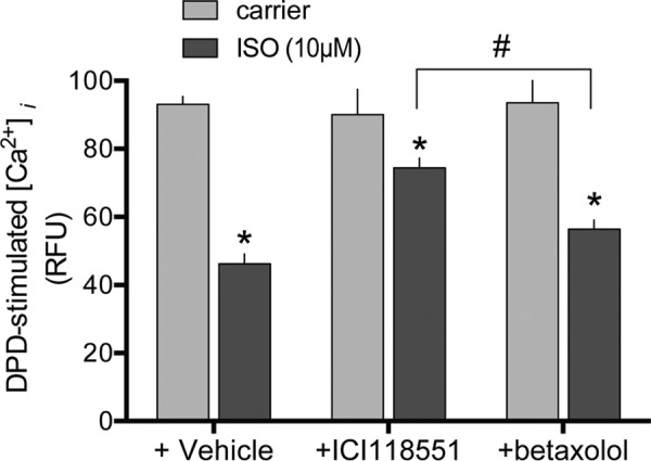
Bitter taste receptors (TAS2Rs) are G-protein-coupled receptors now recognized to be expressed on extraoral cells, including airway smooth muscle (ASM) where they evoke relaxation. TAS2Rs are difficult to express in heterologous systems, with most receptors being trapped intracellularly. We find, however, that co-expression of β2-adrenergic receptors (β2AR) in HEK-293T routes TAS2R14 to the cell surface by forming receptor heterodimers. Cell surface TAS2R14 expression was increased by ∼5-fold when β2AR was co-expressed. Heterodimer formation was shown by co-immunoprecipitation with tagged receptors, biomolecular fluorescence complementation, and merged confocal images. The dynamic nature of this interaction was shown by: a gene-dose relationship between transfected β2AR and TAS2R14 expression, enhanced (up to 3-fold) TAS2R14 agonist stimulation of [Ca(2+)]i with β2AR co-transfection, ∼53% decrease in [Ca(2+)]i signaling with shRNA knockdown of β2AR in H292 cells, and ∼60% loss of [Ca(2+)]i responsiveness in βAR knock-out mouse ASM. Once expressed on the surface, we detected unidirectional, conformation-dependent, interaction within the heterodimer, with β2AR activation rapidly uncoupling TAS2R14 function (∼65% desensitization). Cross-talk was independent of β2AR internalization and cAMP/PKA, and not accompanied by TAS2R14 internalization. With prolonged β-agonist exposure, TAS2R14 internalized, consistent with slow recycling of naked TAS2R14 in the absence of the heterodimeric milieu. In studies of ASM mechanics, rapid cross-talk was confirmed at the physiologic level, where relaxation from TAS2R14 agonist was decreased by ∼50% with β-agonist co-treatment. Thus the β2AR acts as a double-edged sword: increasing TAS2R14 cell surface expression, but when activated by β-agonist, partially offsetting the expression phenotype by direct receptor:receptor desensitization of TAS2R14 function.

Keywords: G protein; asthma; bitter taste receptor; chronic obstructive pulmonary disease (COPD); cyclic AMP (cAMP); dimerization; receptor internalization; smooth muscle.


Figures

FIGURE 1.
FIGURE 1.
Enhanced expression of TAS2R14 by β2AR co-transfection. HEK-293T cells were transfected with the epitope-tagged TAS2R14 construct (see “Experimental Procedures”) in the absence or presence of co-transfection with β2AR (A–E). Cells were exposed to biotin and cell surface proteins were collected using avidin-immobilized agarose beads. The released proteins were subjected to SDS-PAGE and immunoblotted with Myc (A) or FLAG antibody (B). In C, the cell membrane is identified by concanavalin A (green signal) and TAS2R by FLAG antibody (red signal). Merged images reveal TAS2R14 localizes to the cell surface (yellow signal). In D, increased TAS2R14 expression was also observed by confocal imaging (×400), when β2AR was co-transfected. Confocal images (×400) from BiFC studies (E), with the indicated regions of Venus, shows complementation only when they are fused to the two receptors, indicative of TAS2R14-β2AR interaction. Results are representative of 3–5 experiments. Bars: C = 30 μm; D and E = 40 μm.
FIGURE 2.
FIGURE 2.
TAS2R14 and β2AR co-immunoprecipitate and co-localize. HEK-293T cells were transfected with Myc-β2AR and FLAG-TAS2R14 (A). IP with FLAG antibodies followed by SDS-PAGE and immunoblotting (IB) with Myc antibodies only showed a band at the expected Mr when both receptors were transfected. Immunoblotting of the FLAG immunoprecipitates showed signals when FLAG-TAS2R14 was transfected. The inputs to the IP shown in the lower two sections confirm expression of the individual receptors in the cell lysates (10 μg) as indicated. In B, a similar experiment was performed except the IP was with Myc antibody and the IB was with FLAG antibody. For C, co-IP studies with HA- or FLAG-tagged receptors were performed with protein from either membrane fractions (200 μg) or cytosolic fractions (250 μg), and revealed the TAS2R14:β2AR heterodimer in both fractions. Confocal imaging (D, ×600) of HA-β2AR and FLAG-TAS2R14 co-transfected cells shows cell surface expression of each receptor (green and red, respectively) and co-localization (yellow) after merging. Results are representative of 4–6 independent experiments. Bar = 20 μm.
FIGURE 3.
FIGURE 3.
TAS2R14 expressed on HEK-293T cells couple to [Ca2+]i release. Cells transfected with Gα16/G44 with TAS2R14 and β2AR were loaded with Fluo-4 and [Ca2+]i quantitated by fluorescent microscopy (A and B) or by a fluorescence based plate based assay (see “Experimental Procedures”) (C). Control pcDNA-transfected cells showed a minimal to no response to TAS2R agonists (A and B). TAS2R14 + β2AR cells responded to the TAS2R14 agonists quinine (QUI), DPD, and FFA, but not the TAS2R10 agonist strychnine or the TAS2R31 agonist saccharin. *, p < 0.01 versus pcDNA-transfected, n >500 cells imaged per condition. A representative DPD dose-response for stimulation of [Ca2+]i is shown in C. Magnification = ×20, bar = 400 μm.
FIGURE 4.
FIGURE 4.
β2AR expression dynamically regulates TAS2R14 functional expression. HEK-293T cells were transfected with TAS2R14 + Gα16/G44, without or with β2AR (A and B). The [Ca2+]i response to the TAS2R14 agonists DPD and FFA are increased when β2AR is co-transfected, consistent with the increased expression of TAS2R14 (Fig. 1, A and B). In 4 experiments, the [Ca2+]i stimulation was increased by 2.4 ± 0.11 and 2.1 ± 0.12, respectively, when β2AR was co-expressed (p < 0.01 versus absence of β2AR). In C, H292 cells, which endogenously express TAS2R14 and β2AR were transfected with β2AR shRNA (or sh-control) and treated with vehicle or the TAS2R14 agonist DPD. Knockdown of β2AR by β2AR shRNA resulted in decreased TAS2R14-mediated [Ca2+]i signaling. In D and E, βAR knock-out mouse (10) ASM cells (which express no detectable βAR) were challenged with TAS2R agonists and revealed >50% reduction in TAS2R-stimulated [Ca2+]i. Results are from 4 representative experiments.
FIGURE 5.
FIGURE 5.
Activation of β2AR within the β2AR:TAS2R14 heterodimer uncouples TAS2R14 signaling. HASM cells were treated with the β-agonist ISO (10 μm) for 5 min or 1 h, and then cells were challenged with the TAS2R14 agonist DPD (250 μm) with immediate quantitation of [Ca2+]i release (A). Tracings are from a representative experiment performed in triplicate and the inset is mean ± S.E. of 4 experiments. *, p < 0.01 versus vehicle; +, p < 0.05 versus 5 min. In B, cells were treated with ISO or the cAMP stimulator forskolin (FSK, 10 μm) for 5 min and challenged with DPD as in A. Results shown are from a representative experiment of 4 performed, which showed no significant loss (10 ± 8%, p > 0.05) of DPD-stimulated [Ca2+]i from forskolin treatment. In C cells were treated with the agonist ISO (10 μm), the neutral antagonist propranolol (10 μm), or the β2AR inverse agonist ICI118551 (10 μm) for 5 min and then challenged with 250 μm of the TAS2R14 agonist DPD. There was no change in TAS2R14 function with ICI118551 or propranolol pretreatment. #, p < 0.01 versus vehicle, n = 4.
FIGURE 6.
FIGURE 6.
β2AR and TAS2R14 do not co-internalize with ISO exposure. HEK-293T cells co-transfected with HA-β2AR and FLAG-TAS2R14 were imaged by fluorescence microcopy at baseline and after 5 min and 1 h exposure to 10 μm ISO. β2AR (green signal) at 5 min showed a more diffuse receptor signal at the cell surface, and the presence of intracellular receptor signals compared with control, which is prototypical of β2AR internalization. By 1 h, a substantial intracellular β2AR signal is observed with a readily apparent decrease in cell surface expression. In contrast, the distribution of TAS2R14 (red signal) was unchanged at 5 min, consistent with an absence of internalization. But by 1 h of ISO exposure TAS2R14 consistently showed intracellular receptors. Quantitation is shown in Fig. 7A. Images are from a single experiment representative of 5 performed. Magnification = ×600, bar = 30 μm.
FIGURE 7.
FIGURE 7.
Distribution and functional contributions of TAS2R14 internalization by β-agonist. HEK-293T cells were transfected with HA-β2AR and FLAG-TAS2R14. Cells were treated with ISO for 5 min or 1 h, and the change in intracellular expression of each receptor determined by fluorescence microscopy (A) and the change in cell surface expression by the biotinylation assay (see “Experimental Procedures”) (B). As expected, intracellular β2AR increased in a time-dependent manner (A), with concomitant decrease in cell surface expression (B) after 5 min exposure to ISO. In contrast, there was no statistically significant redistribution of TAS2R14 from the cell surface to cytosol at 5 min. However, by 1 h intracellular accumulation and loss of cell surface TAS2R14 was detected. Results are representative of 5 independent experiments, where β2AR cell surface expression loss was 52 ± 2.1 and 86.75 ± 1.1%, and TAS2R14 loss was 11 ± 1.3 and 64 ± 2.6%, at the 5-min and 1-h time points, respectively. In C, the response to TAS2R14 agonist DPD is quantitated after 1 h of ISO in the absence or presence of the internalization inhibitor dynasore. The loss-of-function of TAS2R14 from 1 h ISO was only partially rescued by blocking internalization, consistent with the rapid receptor:receptor uncoupling process still in effect. Results are from a single representative experiment performed in triplicate. See text for statistical analysis from multiple experiments.
FIGURE 8.
FIGURE 8.
Desensitization of adenylyl cyclase activity by TAS2R14 activation. HASM cells were treated with 500 μm FFA for 5 min or 1 h, and then exposed to the indicated cAMP-stimulation agents in the presence of phosphodiesterase inhibition. β2AR (ISO)- and adenylyl cyclase-stimulated (forskolin) cAMP levels were decreased at both time points. When corrected for the decrease in forskolin-stimulated cAMP, β2AR-specific function was found to be unaffected by TAS2R14 agonism (see text). *, p < 0.01 versus vehicle, n = 11 (5 min) or 3 (1 h).
FIGURE 9.
FIGURE 9.
Physiologic consequences of TAS2R:β2AR cross-talk in airway smooth muscle. HASM cells were studied with magnetic twisting cytometry, which measures cell stiffness, where a decrease in stiffness in response to an agonist is the correlate to airway relaxation. In A, cells were treated with 10 μm ISO, 100 μm FFA, or ISO + FFA. All treatment conditions resulted in a decrease in cell stiffness compared with baseline (p < 0.001). However, maximal relaxation was not additive when both agonists were used. The response to the combination was less than when cells were treated with FFA alone. *, response less than FFA alone, p < 0.001. Data shown are results from 104 to 390 individual cell measurements from 3 independent experiments. In B, cells were exposed to FFA in the absence or presence of the neutral βAR antagonist propranolol (10 μm) or the β2AR inverse agonist ICI118551 (10 μm). The FFA-promoted relaxation response was unaffected by either agent. Results are from 231–272 individual cell measurements from 3 independent experiments.
FIGURE 10.
FIGURE 10.
TAS2R14 co-immunoprecipitation studies with other transfected GPCRs. FLAG-TAS2R14 was transfected into HEK293T cells alone or with one of the following HA-tagged GPCRs: β1AR, NMU2R, or CXCR6. Lysates were immunoprecipitated with HA and immunoblotted with FLAG. The upper section shows co-immunoprecipitation of β2AR and β1AR with TAS2R14. No (or very little) co-immunoprecipitation was observed with HA-NUMU2R or HA-CXCR6 transfection. Results are representative of 4 independent experiments.
FIGURE 11.
FIGURE 11.
Effects of β1AR or β2AR activation on TAS2R14 signaling to [Ca2+]i. H1299 cells were pretreated with carrier, the β2AR antagonist ICI118551 (10 μm), or the β1AR antagonist betaxolol (10 μm) for 10 min, and then cells were treated for 5 min with 10 μm ISO. (The latter two conditions selectively activate β1AR and β2AR, respectively.) Desensitization of TAS2R14 signaling was observed with activation of either βAR subtype, and was greater when β2AR was activated compared with β1AR. *, p < 0.01 versus carrier; #, p < 0.01 + ICI118551 versus + betaxolol. Results are from 4 experiments.

References

    1. Chandrashekar J., Hoon M. A., Ryba N. J., and Zuker C. S. (2006) The receptors and cells for mammalian taste. Nature 444, 288–294 - PubMed
    1. Meyerhof W., Batram C., Kuhn C., Brockhoff A., Chudoba E., Bufe B., Appendino G., and Behrens M. (2010) The molecular receptive ranges of human TAS2R bitter taste receptors. Chem. Senses 35, 157–170 - PubMed
    1. Deshpande D. A., Wang W. C., McIlmoyle E. L., Robinett K. S., Schillinger R. M., An S. S., Sham J. S., and Liggett S. B. (2010) Bitter taste receptors on airway smooth muscle bronchodilate by localized calcium signaling and reverse obstruction. Nat. Med. 16, 1299–1304 - PMC - PubMed
    1. Deshpande D. A., Robinett K. S., Wang W. C., Sham J. S., An S. S., and Liggett S. B. (2011) Bronchodilator activity of bitter tastants in human tissue. Nat. Med. 17, 776–778 - PMC - PubMed
    1. Liggett S. B. (2013) Bitter taste receptors on airway smooth muscle as targets for novel bronchodilators. Expert Opin. Therap. Targets 17, 721–731 - PMC - PubMed

Publication types

MeSH terms

Substances