

Psychophysical and rTMS Evidence for the Presence of Motion Opponency in Human V5
- PMID: 27342938
- PMCID: PMC5143189
- DOI: 10.1016/j.brs.2016.05.012
Psychophysical and rTMS Evidence for the Presence of Motion Opponency in Human V5
Abstract
Background: Motion sensitive cells within macaque V5, but not V1, exhibit motion opponency whereby their firing is suppressed by motion in their anti-preferred direction. fMRI studies indicate the presence of motion opponent mechanisms in human V5.
Objective/hypothesis: We tested two hypotheses. 1) Performance of a motion discrimination task would be poorer when stimuli were constructed from pairs of dots that moved in counter-phase vs. in-phase, because counter-phase dots would activate motion opponent mechanisms in V5. 2) Offline 1 Hz rTMS of V5 would impair discrimination performance for in-phase stimuli but not counter-phase stimuli, and the opposite effect would be found for rTMS of V1.
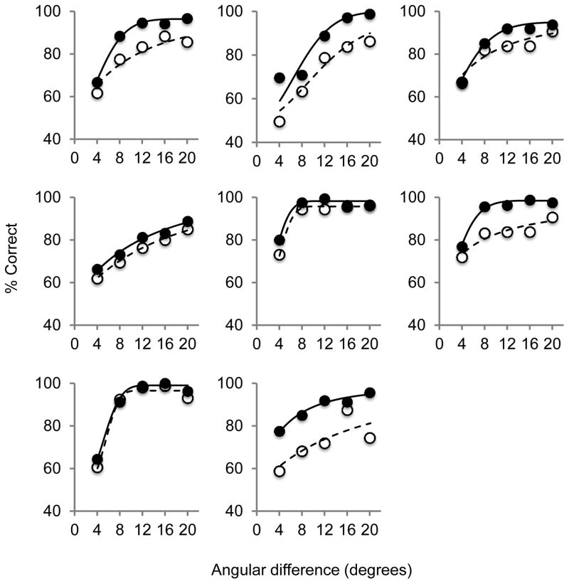
Methods: Stimuli were constructed from 100 dot pairs. Paired dots moved along a fixed motion axis either in counter-phase (motion opponent stimulus) or in-phase (non-opponent motion stimulus). Motion axis orientation discrimination thresholds were measured for each stimulus. Blocks of 300 trials were then presented at 85% correct threshold and discrimination accuracy was measured before and after 1 Hz offline rTMS of either V1 or V5. Subjects were 8 healthy adults.
Results: Discrimination thresholds were significantly larger (worse) for counter-phase than in-phase stimuli (p = 0.02). V5 rTMS mildly impaired discrimination accuracy for the in-phase dot stimuli (p = 0.02) but not the counter-phase dot stimuli. The opposite effect occurred for V1 rTMS (p = 0.05).
Conclusions: Opponent motion mechanisms are present within human V5 and activation of these mechanisms impairs motion discrimination. In addition, perception of the motion axis within opponent motion stimuli involves processing within V1.
Keywords: MT; Middle temporal area; Motion perception; Primary visual cortex; Repetitive transcranial magnetic stimulation; Visual cortex.
Copyright © 2016 Elsevier Inc. All rights reserved.
Figures


Similar articles
-
Spatial proximity modulates the strength of motion opponent suppression elicited by locally paired dot displays.Vision Res. 2018 Mar;144:1-8. doi: 10.1016/j.visres.2018.01.004. Epub 2018 Feb 2. Vision Res. 2018. PMID: 29355566
-
The role of human brain area hMT+ in the perception of global motion investigated with repetitive transcranial magnetic stimulation (rTMS).Brain Stimul. 2015 Mar-Apr;8(2):200-7. doi: 10.1016/j.brs.2014.11.001. Epub 2014 Nov 6. Brain Stimul. 2015. PMID: 25440579 Clinical Trial.
-
The neural basis of form and form-motion integration from static and dynamic translational Glass patterns: A rTMS investigation.Neuroimage. 2017 Aug 15;157:555-560. doi: 10.1016/j.neuroimage.2017.06.036. Epub 2017 Jun 17. Neuroimage. 2017. PMID: 28633972
-
Extrastriate area V5 (MT) and its role in the processing of visual motion.Cesk Fysiol. 2004;53(1):17-22. Cesk Fysiol. 2004. PMID: 15702885 Review.
-
Linking Neuronal Direction Selectivity to Perceptual Decisions About Visual Motion.Annu Rev Vis Sci. 2020 Sep 15;6:335-362. doi: 10.1146/annurev-vision-121219-081816. Annu Rev Vis Sci. 2020. PMID: 32936737 Review.
Cited by
-
Can visual cortex non-invasive brain stimulation improve normal visual function? A systematic review and meta-analysis.Front Neurosci. 2023 Mar 2;17:1119200. doi: 10.3389/fnins.2023.1119200. eCollection 2023. Front Neurosci. 2023. PMID: 36937668 Free PMC article. Review.
-
Effects of cTBS on the Frequency-Following Response and Other Auditory Evoked Potentials.Front Hum Neurosci. 2020 Jul 8;14:250. doi: 10.3389/fnhum.2020.00250. eCollection 2020. Front Hum Neurosci. 2020. PMID: 32733220 Free PMC article.
-
Pathway and directional specificity of Hebbian plasticity in the cortical visual motion processing network.iScience. 2023 Jun 7;26(7):107064. doi: 10.1016/j.isci.2023.107064. eCollection 2023 Jul 21. iScience. 2023. PMID: 37408682 Free PMC article.
References
-
- Orban GA. Higher order visual processing in macaque extrastriate cortex. Physiol Rev. 2008;88:59–89. - PubMed
-
- Born RT, Bradley DC. Structure and function of visual area MT. Annu Rev Neurosci. 2005;28:157–189. - PubMed
-
- De Valois RL, Yund EW, Hepler N. The orientation and direction selectivity of cells in macaque visual cortex. Vision Res. 1982;22:531–544. - PubMed
-
- Schiller PH, Finlay BL, Volman SF. Quantitative studies of single-cell properties in monkey striate cortex. I. Spatiotemporal organization of receptive fields. J Neurophysiol. 1976;39:1288–1319. - PubMed
MeSH terms
Grants and funding
LinkOut - more resources
Full Text Sources
Other Literature Sources
