

Genomic characterization of viral integration sites in HPV-related cancers
- PMID: 27343048
- PMCID: PMC6749823
- DOI: 10.1002/ijc.30243
Genomic characterization of viral integration sites in HPV-related cancers
Abstract
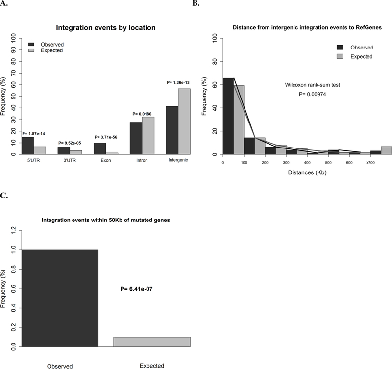
Persistent infection with carcinogenic human papillomaviruses (HPV) causes the majority of anogenital cancers and a subset of head and neck cancers. The HPV genome is frequently found integrated into the host genome of invasive cancers. The mechanisms of how it may promote disease progression are not well understood. Thoroughly characterizing integration events can provide insights into HPV carcinogenesis. Individual studies have reported limited number of integration sites in cell lines and human samples. We performed a systematic review of published integration sites in HPV-related cancers and conducted a pooled analysis to formally test for integration hotspots and genomic features enriched in integration events using data from the Encyclopedia of DNA Elements (ENCODE). Over 1,500 integration sites were reported in the literature, of which 90.8% (N = 1,407) were in human tissues. We found 10 cytobands enriched for integration events, three previously reported ones (3q28, 8q24.21 and 13q22.1) and seven additional ones (2q22.3, 3p14.2, 8q24.22, 14q24.1, 17p11.1, 17q23.1 and 17q23.2). Cervical infections with HPV18 were more likely to have breakpoints in 8q24.21 (p = 7.68 × 10(-4) ) than those with HPV16. Overall, integration sites were more likely to be in gene regions than expected by chance (p = 6.93 × 10(-9) ). They were also significantly closer to CpG regions, fragile sites, transcriptionally active regions and enhancers. Few integration events occurred within 50 Kb of known cervical cancer driver genes. This suggests that HPV integrates in accessible regions of the genome, preferentially genes and enhancers, which may affect the expression of target genes.
Keywords: HPV; integration.
© 2016 UICC.
Conflict of interest statement
The authors have no conflicts of interest.
Figures


References
-
- Schiffman M, Castle PE, Jeronimo J, Rodriguez AC, Wacholder S. Human papillomavirus and cervical cancer. Lancet 2007;370: 890–907. - PubMed
-
- Madsen BS, Jensen HL, van den Brule AJ, Wohlfahrt J, Frisch M. Risk factors for invasive squamous cell carcinoma of the vulva and vagina--population-based case-control study in Denmark. Int J Cancer 2008;122: 2827–34. - PubMed
-
- Klaes R, Woerner SM, Ridder R, Wentzensen N, Duerst M, Schneider A, Lotz B, Melsheimer P, von Knebel Doeberitz M. Detection of high-risk cervical intraepithelial neoplasia and cervical cancer by amplification of transcripts derived from integrated papillomavirus oncogenes. Cancer Res 1999;59: 6132–6. - PubMed
Publication types
MeSH terms
Grants and funding
LinkOut - more resources
Full Text Sources
Other Literature Sources
Medical
