. 2016 Aug 1;311(2):L389-99.
doi: 10.1152/ajplung.00171.2016. Epub 2016 Jun 24.

Dose dependency of outcomes of intrapleural fibrinolytic therapy in new rabbit empyema models

Affiliations

Dose dependency of outcomes of intrapleural fibrinolytic therapy in new rabbit empyema models

Andrey A Komissarov et al. Am J Physiol Lung Cell Mol Physiol. .

Abstract

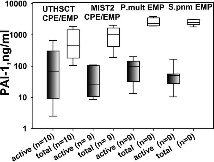
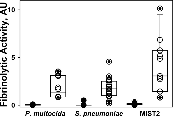
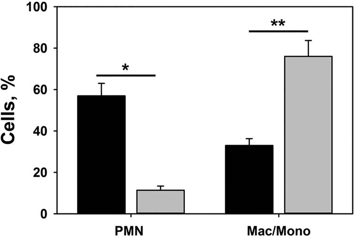
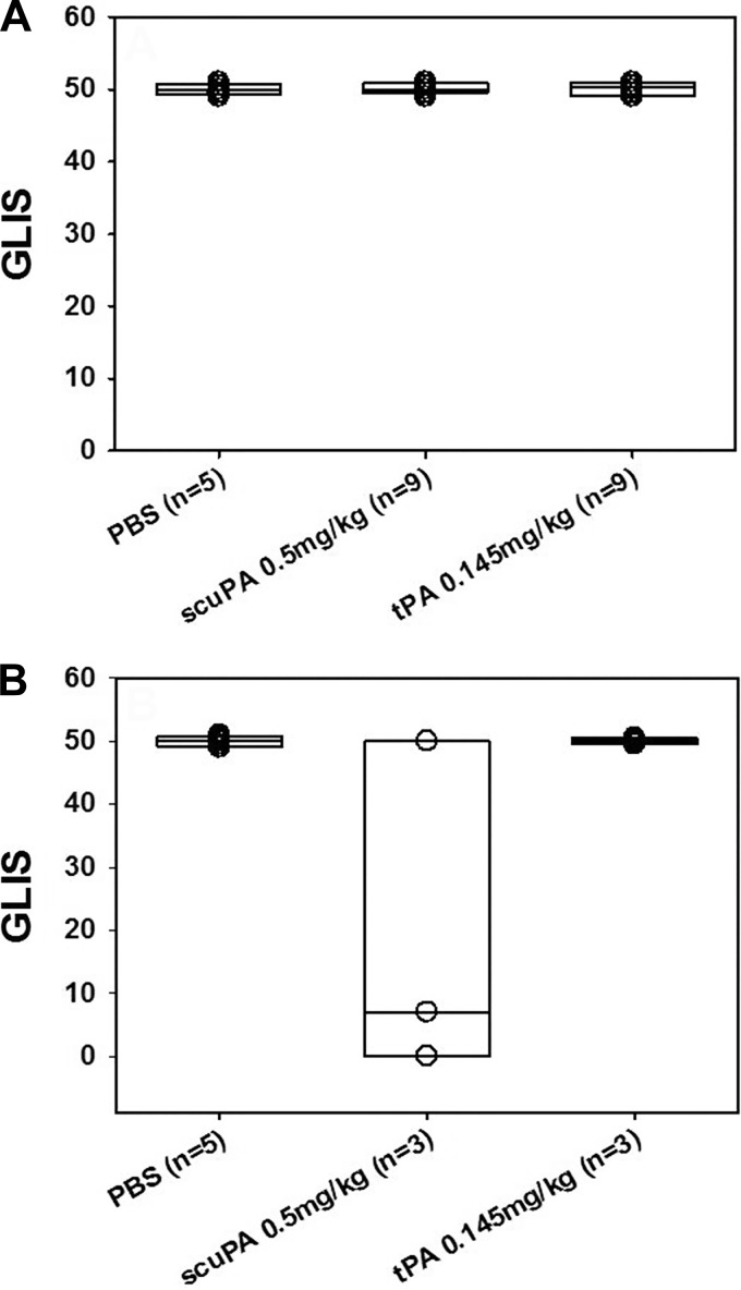
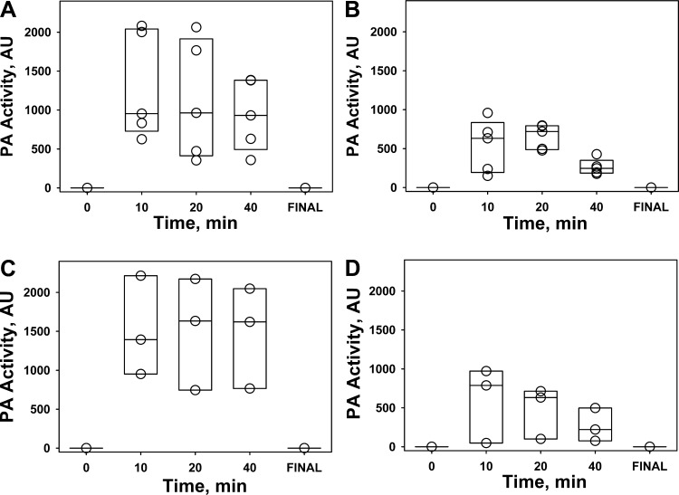
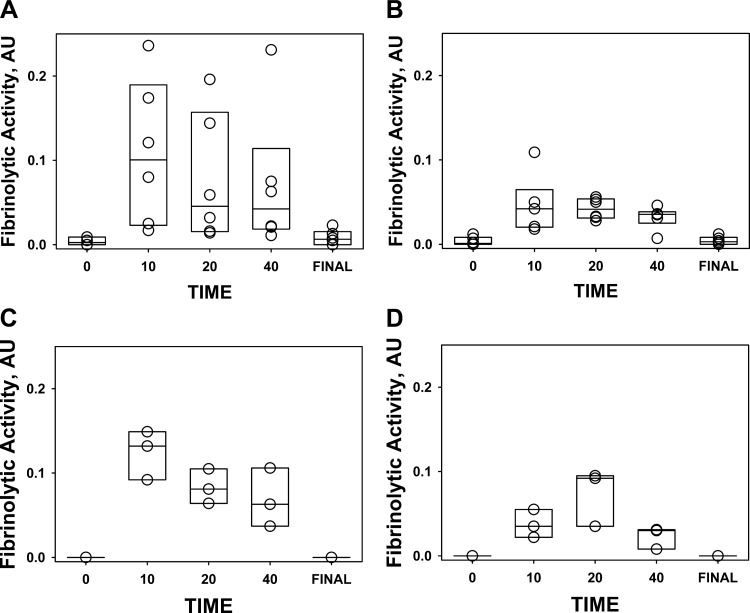
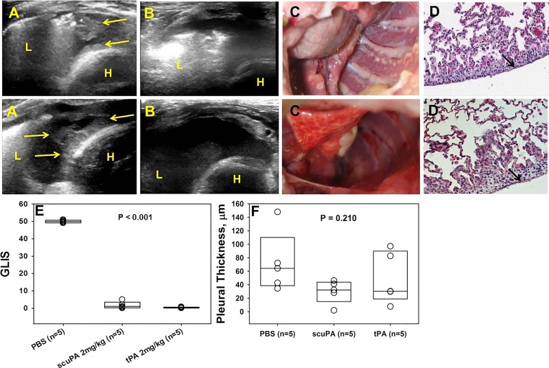
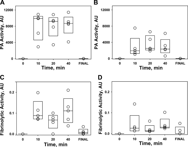
The incidence of empyema (EMP) is increasing worldwide; EMP generally occurs with pleural loculation and impaired drainage is often treated with intrapleural fibrinolytic therapy (IPFT) or surgery. A number of IPFT options are used clinically with empiric dosing and variable outcomes in adults. To evaluate mechanisms governing intrapleural fibrinolysis and disease outcomes, models of Pasteurella multocida and Streptococcus pneumoniae were generated in rabbits and the animals were treated with either human tissue (tPA) plasminogen activator or prourokinase (scuPA). Rabbit EMP was characterized by the development of pleural adhesions detectable by chest ultrasonography and fibrinous coating of the pleura. Similar to human EMP, rabbits with EMP accumulated sizable, 20- to 40-ml fibrinopurulent pleural effusions associated with extensive intrapleural organization, significantly increased pleural thickness, suppression of fibrinolytic and plasminogen-activating activities, and accumulation of high levels of plasminogen activator inhibitor 1, plasminogen, and extracellular DNA. IPFT with tPA (0.145 mg/kg) or scuPA (0.5 mg/kg) was ineffective in rabbit EMP (n = 9 and 3 for P. multocida and S. pneumoniae, respectively); 2 mg/kg tPA or scuPA IPFT (n = 5) effectively cleared S. pneumoniae-induced EMP collections in 24 h with no bleeding observed. Although intrapleural fibrinolytic activity for up to 40 min after IPFT was similar for effective and ineffective doses of fibrinolysin, it was lower for tPA than for scuPA treatments. These results demonstrate similarities between rabbit and human EMP, the importance of pleural fluid PAI-1 activity, and levels of plasminogen in the regulation of intrapleural fibrinolysis and illustrate the dose dependency of IPFT outcomes in EMP.

Keywords: empyema; fibrinolysis; plasminogen activator inhibitor-1; single chain urokinase; tissue plasminogen activator.


Figures

Fig. 1.
Fig. 1.
Pleural injury in acute P. multocida and S. pneumoniae EMP in rabbits. Top: chest ultrasonography (A) at 96 h after induction of EMP; yellow arrows indicate pleural adhesions seen as rounded opaque structures. L, lung; H, heart. Postmortem visual evaluation (B) shows extensive adhesions (black arrows) with fibrinous coating of the lung. Bottom: histology of pleural injury. Tissue sections were prepared from the lungs of rabbits with P. multocida and S. pneumoniae-induced pleural injury (96 h after induction of the model), n = 3 per group. Tissue sections were stained with hematoxylin and eosin (H&E) and probed for collagen via trichrome stain and fibrin via immunohistochemistry. The entire sectioned pleura was analyzed for fibrin (red) and collagen (blue) deposition. Arrows indicate fibrin deposition. All images were taken at ×20 magnification on a Nikon Eclipse Ti inverted microscope. Illustrated findings are representative of 3 sections/group. Averaged pleural thickness (surface to basement membrane) was 580 and 60 μm for P. multocida and S. pneumoniae, respectively, as determined by morphometry.
Fig. 2.
Fig. 2.
Both active and total PAI-1 levels at baseline pleural fluids from rabbits with EMP are similar to those observed in human EMP. A semilogarithmic plot of concentration (ng/ml) of active (shaded) and total (open) PAI-1 in samples of EMP/complicated pleural effusion (CPE) pleural fluid (from left to right) from humans prospectively collected at UTHSCT; patients from the participating in the MIST2 clinical trial; rabbits with P. multocida (P.mult) and S. pneumoniae (S.pnm)-induced EMP, respectively. Commercially available ELISAs (Molecular Innovation) were used. Data are presented as box plots (showing interquartile ranges). Results for Kruskal-Wallis test demonstrated no statistically significant difference (P > 0.05) in the medians either for active PAI-1 or for total antigen among the 4 groups.
Fig. 3.
Fig. 3.
Activation of endogenous plasminogen results in a significant increase in the suppressed fibrinolytic activity in baseline pleural fluids from rabbits with P. multocida and S. pneumoniae EMP and humans from the MIST2 clinical trials. Aliquots of pleural fluid withdrawn prior to IPFT were analyzed by FITC-fibrin film fibrinolytic activity assay (26) with (○) or without (●) supplementation with 5 nM of 2-chain urokinase (tcuPA) (13). A significant increase in the baseline fibrinolytic activity occurred after supplementation with exogenous fibrinolysin (○) as observed in pleural fluid samples of (from left to right) rabbits with P. multocida and S. pneumoniae-induced EMP and humans from the MIST2 trials.
Fig. 4.
Fig. 4.
Extracellular DNA concentration in pleural fluids of humans and rabbits with EMP. A: semilogarithmic plot of DNA concentration (μg/ml) in human (prospectively collected at UTHSCT and MIST2) and rabbit P. multocida and S. pneumoniae-induced EMP pleural fluids. Total nucleic acids were purified from 100 μl of pleural fluid by using a DNeasy blood and tissue kit from Qiagen as described in the manufacturer's protocol. The amounts and size distribution of DNA were estimated by A260 (A) and agarose gel electrophoresis (B). Lane 1, Human UTHSCT; lane 2, human MIST2; lane 3, rabbit with P. multocida EMP; lane 4, rabbit with S. pneumoniae EMP. The reassembly of noncontiguous gel lanes is demarcated by white spaces. Gels were visualized and analyzed by using a Molecular Imager supplemented with Quantity One (version 4.2.3) software (Bio-Rad). Data are presented as box plots (showing interquartile ranges). Results for Kruskal-Wallis test presented statistically significant difference in the mean values among the treatment groups (P < 0.001). Pairwise multiple comparison showed that the concentration of DNA in the P. multocida pleural injury group is statistically higher among the analyzed groups (P < 0.001).
Fig. 5.
Fig. 5.
Pleural fluid WBC differential counts in rabbit P. multocida and S. pneumoniae-induced pleural injury. Pleural fluids were collected at the time of euthanasia and RBC and WBC count was performed as described in materials and methods. A predominance of neutrophils (PMN) is noted in S. pneumoniae injury whereas a mixed myeloid cell distribution was seen at 96 h after induction of P. multocida-induced EMP. Results of paired t-tests showed statistically significant change P = 0.013 (*) and 0.006 (**) in cell populations. Data are presented as a grouped vertical bar graph. Mac/Mono, macrophages/monocytes.
Fig. 6.
Fig. 6.
IPFT with MED of tPA or scuPA identified for TCN-induced rabbit pleural injury (18, 24) was ineffective in treatment of P. multocida (A) and S. pneumoniae (B) rabbit EMP. A: outcomes of IPFT of rabbits with P. multocida EMP expressed as GLIS scores (23, 25) for treatment (from left to right) with the vehicle (n = 5), scuPA (0.5 mg/kg; n = 9), and tPA (0.145 mg/kg; n = 9). Data are presented as box plots (showing interquartile ranges). Results for Kruskal-Wallis tests showed no statistically significant difference (P > 0.05) in medians among the treatment groups. B: outcomes of IPFT of rabbits with EMP for treatment (from left to right) with vehicle (n = 5), scuPA (0.5 mg/kg; n = 3) and tPA (0.145 mg/kg; n = 3). Data are presented as box plots (showing interquartile ranges). Kruskal-Wallis tests showed no statistically significant difference (P > 0.05) in medians among the treatment groups.
Fig. 7.
Fig. 7.
Unsuccessful IPFT of acute P. multocida (top) and S. pneumoniae (bottom) EMP with MED of scuPA or tPA identified for TCN-induced pleural injury (18, 24). Rabbits with acute EMP were treated with 0.5 mg/kg of scuPA or 0.145 mg/kg tPA at 72 h (Fig. 3). A: pleural injury was evaluated at 96 h (24 h after IPFT) by gross examination (arrows indicate fibrin depositions and lung coating) and gross findings were confirmed by microscopic examination and by immunostaining that showed fibrin (red) on the visceral pleural surface (B). All images were taken at ×20 magnification on a Nikon Eclipse Ti inverted microscope. Illustrated findings are representative of 3 sections/group. C: pleural thickness (surface to basement membrane) was determined by morphometry (30 fields/slide, n = 3 representative sections per animal). Data are presented as box plots (showing interquartile ranges). Kruskal-Wallis tests showed no statistically significant difference (P > 0.05) in medians among the treatment groups.
Fig. 8.
Fig. 8.
Changes in intrapleural PA activity during IPFT of rabbits with P. multocida (A and B) and S. pneumoniae (C and D) EMP with scuPA (0.5 mg/kg; A and C) and tPA (0.145 mg/kg; B and D). Changes in enzymatic PA activity in pleural fluids withdrawn at 10, 20, and 40 min and at 24 h (Final) represent kinetics of intrapleural inactivation of scuPA (A and B) and tPA (C and D). PA activity was measured as described in materials and methods and elsewhere (13, 24).
Fig. 9.
Fig. 9.
Changes in the fibrinolytic activity in pleural fluids of rabbits with P. multocida (A and B) and S. pneumoniae (C and D) EMP during IPFT with scuPA (0.5 mg/kg; A and C) and tPA (0.145 mg/kg; B and D). Fibrinolytic activity in pleural fluids withdrawn at 0, 10, 20, and 40 min and at the end of the experiment (Final) after injection 0.5 mg/kg (n = 3) scuPA (A, n = 5; C, n = 3) or 0.145 mg/kg tPA (B, n = 5; D, n = 3). Fibrinolytic activity in aliquots of pleural fluid was measured with a FITC-fibrin film assay as described previously (26).
Fig. 10.
Fig. 10.
Effective IPFT of S. pneumoniae EMP in rabbits with 2 mg/kg of tPA or scuPA clears intrapleural fibrinous adhesions and pleural rind. Rabbits with acute S. pneumoniae-induced EMP were treated with 2 mg/kg of scuPA (top) or tPA (bottom) at 72 h and euthanized at 24 h after. A: chest ultrasonography immediately before IPFT demonstrated extensive opaque pleural adhesions and fibrin deposition (yellow arrows, L, lung; H, heart). B: chest ultrasonography 8-h IPFT demonstrated dissolution of pleural adhesions and fibrin deposition. Pleural effusion and adhesions were cleared 24 h after IPFT as seen on gross examination (C); this was confirmed by immunostaining, which showed very little, focal fibrin (red) deposition on the visceral pleural surface (D). All images were taken at ×40 magnification on a Nikon Eclipse Ti inverted microscope. Illustrated findings are representative of 3 sections/group. E: outcomes of IPFT were expressed as GLIS [GLIS < 10 corresponds to effective IPFT (13, 25)] for treatment with the vehicle (n = 5), scuPA 2 mg/kg (n = 5), and tPA 2 mg/kg (n = 5). Data are presented as box plots showing interquartile ranges. Kruskal-Wallis tests showed statistically significant differences (P < 0.001) in medians among the treatment groups. F: pleural thickening (surface to basement membrane) was determined by morphometry (30 fields/slide, n = 3 representative sections per animal). Kruskal-Wallis tests did not show statistically significant difference in medians among the treatment groups (P = 0.210).
Fig. 11.
Fig. 11.
Changes in intrapleural PA (A and B) and fibrinolytic (C and D) activities during IPFT of S. pneumoniae EMP in rabbits with 2 mg/kg of scuPA (A and C) and tPA (B and D). PA and fibrinolytic activities were determined in pleural fluids of rabbits with S. pneumoniae-induced EMP withdrawn at 0, 10, 20, and 40 min and at the end of the experiment (24 h, final) after injection 2.0 mg/kg of scuPA (n = 3; A and C) or tPA (n = 3; B and D). Changes in PA and fibrinolytic activities in pleural fluids withdrawn at the indicated time were measured as described in materials and methods (13, 24).

References

    1. Abu-Daff S, Maziak DE, Alshehab D, Threader J, Ivanovic J, Deslaurier V, Villeneuve PJ, Gilbert S, Sundaresan S, Shamji F, Lougheed C, Seely JM, Seely AJ. Intrapleural fibrinolytic therapy (IPFT) in loculated pleural effusions—analysis of predictors for failure of therapy and bleeding: a cohort study. BMJ Open 3: 2013. - PMC - PubMed
    1. Asselbergs FW, Williams SM, Hebert PR, Coffey CS, Hillege HL, Navis G, Vaughan DE, van Gilst WH, Moore JH. The gender-specific role of polymorphisms from the fibrinolytic, renin-angiotensin, and bradykinin systems in determining plasma t-PA and PAI-1 levels. Thromb Haemost 96: 471–477, 2006. - PubMed
    1. Bouros D, Antoniou KM, Chalkiadakis G, Drositis J, Petrakis I, Siafakas N. The role of video-assisted thoracoscopic surgery in the treatment of parapneumonic empyema after the failure of fibrinolytics. Surg Endosc 16: 151–154, 2002. - PubMed
    1. Cesari M, Pahor M, Incalzi RA. Plasminogen activator inhibitor-1 (PAI-1): a key factor linking fibrinolysis and age-related subclinical and clinical conditions. Cardiovasc Ther 28: e72–e91, 2010. - PMC - PubMed
    1. Chiu CY, Wong KS, Huang JL, Tasi MH, Lin TY, Hsieh SY. Proinflammatory cytokines, fibrinolytic system enzymes, and biochemical indices in children with infectious para-pneumonic effusions. Pediatr Infect Dis J 27: 699–703, 2008. - PubMed

Publication types

MeSH terms